| MEDICAL INTRO |
| BOOKS ON OLD MEDICAL TREATMENTS AND REMEDIES |
THE PRACTICAL |
ALCOHOL AND THE HUMAN BODY In fact alcohol was known to be a poison, and considered quite dangerous. Something modern medicine now agrees with. This was known circa 1907. A very impressive scientific book on the subject. |
DISEASES OF THE SKIN is a massive book on skin diseases from 1914. Don't be feint hearted though, it's loaded with photos that I found disturbing. |
170 INFLAMMATIONS
GRANULOMA ANNULARE
Synonyms.—Ringed eruption on the fingers (Colcott Fox); Lichen annularis,
Ringed eruption of the extremities (Galloway); Sarcoid tumors (Rasch, Galewski);
Eruption chronique circinée de la main (Dubreuilh); Neoplasie nodulaire et circinée
(Brocq); Erythematosclerosis circinée du dos des mains (Audry).
Granuloma annulare is a rare chronic dermatosis observed more
commonly in children, and most frequently on the dorsal aspect of the
hands, especially over the joints; and consisting usually of several some
what deep-seated and projecting whitish or pinkish nodules and con
tinuous or broken whitish nodular rings. This peculiar and interesting
malady has been given established recognition through the observations
of Colcott Fox, Dubreuilh, Galloway, Crocker, Brocq, Graham Little,
and others.1
Symptoms.—The malady may present itself somewhat suddenly,
but usually gradually and slowly; and it may begin as one or several
1 Literature: Colcott Fox, “Ringed Eruption on the Fingers,” Brit. Jour. Derm.,
1895, p. 91 (case demonstration), and “Ringed Nodular Eruption,” ibid., 1896, p. 15
(case demonstration); Dubreuilh, “Sur un cas d‘Eruption circinée chronique de la
main,” Annales, 1895, p. 355 (with histologic examination), and ibid., 1905, p. 65
(3 additional cases); Galloway, “Lichen Annularis: A Ringed Eruption of the Ex
tremities,” Brit. Jour. Derm., 1899, p. 221 (with excellent colored plate, two histologic
cuts, and review of similar and allied cases, with references); Crocker, “Granuloma
Annulare,” ibid., 1902, p. 1 (6 cases: 4 personal, 1 Pringle‘s, 1 Pernet‘s; colored plate,
showing 3 cases and histologic cuts); Brocq, Annales, 1904, p. 1089, “Neoplasié nodu-
laire et circinée des extremités,” and “Traité elementaire de dermatologie pratique,”
vol. ii, p. 275 (2 case illustrations); Galewski, Iconographia Dermatologica, Fasc iii
(colored plate); Graham Little, “Granuloma Annulare,” Brit. Jour. Derm., 1908, pp.
213, 248, 281, and 317, in his excellent and exhaustive paper, gives a résumé and
references of the above and all other published cases, and of a number of communi
cated (unpublished) cases, with illustrations of the Galloway, Sequeira, Leslie Roberts,
Hyde and Montgomery, Macleod, Colcott Fox, and his own cases; and histologic cuts
of the Pernet, Pringle, Whitfield, Savill, Jadassohn, Adamson, and his own cases; and
an analytic tabulation of 49 cases; discussion of this paper by Crocker, Galloway,
Pernet, Colcott Fox, and Adamson, ibid., p. 327; Crocker, Jour. Cutan. Dis., 1894, p. 5
(reported as a case of lupus erythematosus resembling lichen planus); Pernet, case of
granuloma annulare (celluloma annulare, Pernet) (with illustrations), Proceedings of
the Royal Society of Medicine, London, 1908; G. W. Wende, “A Nodular, Terminating
in a Ring, Eruption (Granuloma Annulare),” Jour. Cutan. Dis., 1909, p. 388 (case
illustrated and histologic cuts); dalla Favera, Dermatolog. Zeitschr., 1909, vol. xvi, p. 73
(case and histologic illustrations; first Italian case); Halle (Lesser's clinic), Archiv,
1909, vol. xcix, p. 51 (report of a case, with review, case and histologic illustrations
[colored]); Hartzell, Jour. Cutan. Dis., 1910, p. 302 (case demonstration; x-ray
exposures had already flattened the lesions considerably; Pellier, “Stereo-phlogose
nodulaire et circinée (Granuloma annulare de Crocker”), Annales, 1910, p. 28; on
hand; Graham Little, Brit. Jour. Derm., 1910, p. 390 (case demonstration); Varney
and Jamieson, Jour. Cutan. Dis., 1911, p. 22, illustration, male patient, aged fifty-eight,
lesions on wrist and hand, gradually disappeared under arsenic; MacLeod, Brit. Jour.
Derm., 1911, p. 409 (case demonstration), girl aged four; on back of both thighs and
calves; Bunch, Brit. Jour. Derm., 1911, p. 357 (case demonstration), boy aged two
and one-half years, on dorsum of right foot; Chipman, Brit. Jour. Derm., Nov., 1911,
p. 349, boy aged fourteen; on pinna of each ear and back of each hand (case and histo-
logic illustrations); C. J. White, Boston Med. and Surg. Jour., May 4, 1911; girl aged
eight, index fingers; gradually disappeared under x-ray exposures (histologic ex
amination); Vignolo-Lutati, Dermatolog. Wochenschr., Jan. 20 and 27,1912,pp. 77 and
114; girl aged thirteen, on dorsum of hand—disappeared on administration of sodium
salicylate, leaving a small atrophic scar; histologic study; careful review of the litera
ture; Piccardi, “Erythema Elevatum et diutinum,” Dermatolog. Wochenschr., Sept. 7,
1912, vol. lv, p. 1115, review and bibliography; discussion of the two conditions—
erythema elevatum and granuloma annulare.
GRANULOMA ANNULARE
171
discrete nodules, as a more or less ringed or crescentic group of nod
ules, or possibly (?) as a distinct continuous ring. The formation is
seemingly semitranslucent, has a smooth surface, is whitish or ivory-
like, often shining and glistening in appearance; sometimes with a bluish-
red or purplish-red tinge which is occasionally quite pronounced and
somewhat deep in hue. It is a solid formation, either firm or slightly
doughy to the touch; deeply seated as well as projecting above the
skin level, with, as a rule, a narrow areolar pinkish or reddish zone. It
is usually a trifle flattened or it may be distinctly so. A beginning
nodule increases in size to that of a small to large pea, and may remain
as such; but it may increase peripherally in area and with a partial or
complete disappearance of the central part, finally presenting as a per
fectly or imperfectly formed elevated ring-like or crescentic plaque, the
band being 1/16 to 1/8 inch, or occasionally more in width. When begin
ning as a ringed or crescentic group of nodules, these enlarge, crowd
together more or less closely at the contiguous sides, with a resulting
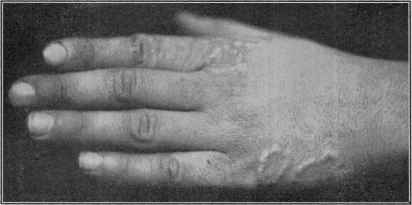
Fig. 30.—Granuloma annulare.
ring-like plaque. A plaque may be artistically ring-like, or it may only
be irregularly and unevenly circular or crescentic in outline; not infre
quently a small or even large portion of the ring missing, the resulting
plaque being a crescent or a segment of a ring. The ring, though upon
casual inspection occasionally appearing solid and continuous, is rarely
unbroken, but is made up of contiguous or closely set, sometimes fused,
nodules.
The size of the ring-like formation varies from a fraction of an inch
to 1 or 2 inches or more in diameter. The skin of the inclosed area
seems normal, but upon inspection with a lens slight atrophy may be
observed in some instances; it may be the normal skin color or pinkish
or reddish. Its course is usually persistent, after an uncertain develop
ment, often remaining stationary for some time or almost indefinitely,1
sometimes one or more of the lesions partly disappearing or entirely
1 Colcott Fox (discussion on Dr. Bunch's case, loc. cit.) mentioned a case in a
woman he saw twenty years ago, and in whom it still continues.
172
INFLAMMATIONS
disappearing, with now and then a new nodule or ring presenting.
Doubtless, in most instances, after an uncertain period of several months
or years, it undergoes spontaneous involution and cure, slight stains
marking the sites for a time.1 There are no subjective symptoms, only
rarely is slight evanescent burning, itching, or tenderness complained of.
The eruption is seldom abundant, usually consisting of not more than
several nodules and rings; most cases are only seen after the ring forma
tion or grouping is more or less fully developed. The most common site
for the malady is the dorsal surface of the hands, especially over the
joints; next in frequency, in the order named, are wrists, feet, ankles,
neck, elbows, knees, and buttocks; face and scalp are rarely affected
(Graham Little).
Etiology and Pathology.—The cause of the disease is not
known; it is thought to occur more frequently in those of tuberculous
antecedents. It is more commonly observed in children and early
youth, and about equally in the two sexes. In a number of instances
it first presented in summer time. The histologic conditions, studied
by most observers named, do not justify the term “granuloma.” Gallo
way found the process to be an inflammatory one, consisting chiefly of
cell infiltration of the type seen in certain chronic inflammatory processes
in the cutis, especially the lichen group. Graham Little concludes that
we have to do with a deep hypodermic inflammation gradually spread
ing toward the surface, and situated around vessels; the cell masses, con
sisting of large mononuclear cells, numerous spindle-shaped, or oblong,
or pear-shaped cells, with an elongated nucleus, indistinguishable from
connective-tissue corpuscles; and a few large epithelioid cells inter
spersed in the cell mass; in many of the foci of cells there appeared to be
central destruction; there were no plasma cells, and only occasionally
mast cells in abnormal numbers.
Diagnosis.—The peculiar whitish or ivory-colored nodule, ele
vated band-like or nodular rings, segments or crescents, its sluggish
course, and the absence of subjective symptoms are so distinctive that
the malady can scarcely be confounded with anything else. Lichen
planus annularis bears slight resemblance, and some observers claim
relationship with erythema elevatum diutinum. Exceptionally it has
some keloidal suggestion.
Prognosis and Treatment—The malady is benign, finally, after
a variable period of months or years, probably disappearing sponta
neously. Sodium salicylate (Vignolo-Lutati) and arsenic (Varney and
Jamieson) have been credited with favorable influences. As a rule, the
lesions will yield more or less rapidly to applications which tend to pro
duce desiccation and exfoliation; salicylic acid and resorcin ointments,
pastes, lotions or paints, such as are employed in acne, callosity, and
senile keratoses. X-ray has been favorably spoken of (Hartzell, C. J.
White).
1 Graham Little, Brit. Jour. Derm., 1912, p. 22 (case demonstration), notes a recur
rence in patient previously under his care, after a few years’ freedom.
But first, if you want to come back to this web site again, just add it to your bookmarks or favorites now! Then you'll find it easy!
Also, please consider sharing our helpful website with your online friends.
BELOW ARE OUR OTHER HEALTH WEB SITES: |
Copyright © 2000-present Donald Urquhart. All Rights Reserved. All universal rights reserved. Designated trademarks and brands are the property of their respective owners. Use of this Web site constitutes acceptance of our legal disclaimer. | Contact Us | Privacy Policy | About Us |