| MEDICAL INTRO |
| BOOKS ON OLD MEDICAL TREATMENTS AND REMEDIES |
THE PRACTICAL |
ALCOHOL AND THE HUMAN BODY In fact alcohol was known to be a poison, and considered quite dangerous. Something modern medicine now agrees with. This was known circa 1907. A very impressive scientific book on the subject. |
DISEASES OF THE SKIN is a massive book on skin diseases from 1914. Don't be feint hearted though, it's loaded with photos that I found disturbing. |
HERPES ZOSTER
Synonyms.—Zona; Zoster; Shingles; Ignis sacer; Fr., Zona; Ger., Gürtelaus-
schlag; Feuergürtel.
Definition.—An acute inflammatory self-limited disease, char
acterized by the appearance of several or more groups of vesicles on
slightly elevated and inflamed areas, of unilateral distribution, and
corresponding to the peripheral and intertwining branches of one or
two cutaneous nerves.
Symptoms.—In many instances there is more or less neuralgic
pain in the region for one or several days preceding the cutaneous lesions.
This may continue through the course of the disease, being continuous
or intermittent in character, or it may abate when the eruption is fully
out. In other cases the outbreak of the vesicles and neuralgic pains are
synchronous. Not infrequently the pain may be so slight as to give rise
to no complaint, or sometimes it is entirely absent; this is observed more
especially in children (Bohn). In some of the more extensive cases there
may be, in the beginning, mild febrile action, chilliness, and a variable
degree of malaise. Swelling of the neighboring, and occasionally other,
lymphatic glands is frequently, and probably always, noted (Barthélemy,
Strümpfell, Blaschko, Winfield, Hay, and others).1 The eruption
1 Barthélemy, Annales, 1891, p. 21, and 1892, p. 168.
HERPES ZOSTER
349
makes its appearance suddenly, usually as several or more hyperemic
or slightly inflammatory patches, upon which are seated usually 10
to 20 papules or vesicopapules, irregularly grouped; these, generally
before the cases is seen by the physician, soon become clearly defined
vesicles, of the size varying from a pin-head to a pea; two or three closely
crowded together sometimes become confluent, and form a bean-sized
bleb. They show no tendency to spontaneous rupture. New vesicles
and patches may come out for several days or longer, although in most
cases all the patches are concomitant or are out within forty-eight hours.
The disease reaches its full development in five to ten days, and then be-
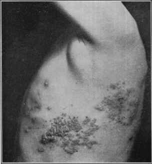
Fig. 82.—Herpes zoster (dorsopectoralis), left pectoral region, in a youth of sixteen,
of about one week‘s duration. The grouping and cluster tendency are shown; a few
lesions in the patch on the side slightly hemorrhagic There were also a few groups on
the same level posteriorly.
gins to subside. The contents of the lesion are clear, becoming slightly
milky, and rarely puriform; at the end of one to two weeks they have
dried to thin, yellowish or brownish crusts, which in several days drop
off, leaving red spots, which gradually fade, in most instances no perma
nent trace remaining. Sometimes, however, there may be a variable
amount of scarring left to mark the site of the vesicles. Occasionally
the eruption does not go beyond the vesicopapular stage (abortive zoster).
In some cases, more especially in old people and in those in a depraved
condition of health, the lesions, or some of them are hemorrhagic (herpes
zoster hæmorrhagicus), and contain an admixture of blood and pus;
in other exceptional instances there is a slight or marked degree of gan-
350
INFLAMMATIONS
grenous action (herpes zoster gangrænosus),1 and several such gangrenous
vesicles may coalesce, producing areas of ulceration, usually superficial
in character, but which may finally result in considerable scarring. In
this latter class of cases, especially, there is, as a rule, more or less con
stitutional disturbance of fever, loss of appetite, nausea, and chilliness,
and in rare instances the patient becomes septicemic and succumbs.
Exceptionally lymphangitis, furuncles, carbuncles, and phlegmon have
been noted as complications (Besnier and Doyon, von Düring).
There is frequently a sense of soreness or burning at the seat of the
malady, and exceptionally itching. The neuralgic pain may in some in
stances continue long after the complete disappearance of the lesions.
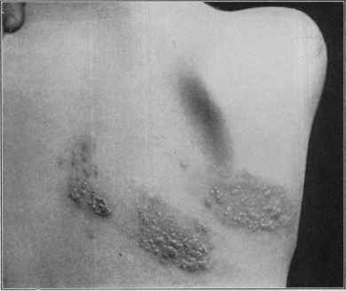
Fig. 83.—Herpes zoster (dorsopectoralis) of right pectoral region, in a male adult,
of about five days’ duration, showing the erythematous plaques with the numerous vesi
cles, some coalescent. A few patches also anteriorly (courtesy of Dr. M. B. Hartzell).
The eruption may appear upon any portion of the body, following the
course of a nerve or of two or more nerves; it is, therefore, always limited
in extent, in some cases, however, much more extensive than in others;
exceptionally, however, it has been noted to involve a greater part
(Wetherill) or the entire half of the trunk (Wilson), several or more nerves
being implicated. In slight cases, on the other hand, there may not be
more than two or three small groups. With rare exceptions the eruption
is unilateral, with, in rare instances, a few lesions seen at a distance from
the seat of the disease (Jamieson, Girandeau, Jeanselme, and Leredde).
1 Baum, “Herpes Zoster Gangrænosus,” Medicine, 1895, p. 1 (with colored plate),
describes a case, and refers briefly to Kaposi‘s and other similar instances.
Plate XI.
Herpes zoster.
HERPES ZOSTER
351
Sometimes one or two groups are seen just beyond or overstepping the
median line. Exceptionally it is bilateral (Moers, Bryant, Squire,
Stabell, Finny, Elliot, Carpenter, Hallopeau et Barrie, Colcott Fox,
and others),1 and usually on the same plane, but cases have also been
observed when the eruption on one side (usually chest or abdomen) was
higher or lower than that on the other. These exceptions to the unilateral
distribution are, however, extremely rare, many observers, including my
self, of large clinical opportunities never having met with a single instance.
In very rare instances, in addition to the characteristic unilateral
zoster eruption of a region, there appear along with it discrete and
scattered, small to large pea-sized, vesicles over the general surface,
usually somewhat scanty in number.2
The most common sites for herpes zoster are the thoracic, lumbar,
and supra-orbital regions. Various regional names are given to the
malady, simply to indicate the locality on which the eruption occurs.
The principal of these are zoster capillitti, zoster faciei, zoster ophthal-
micus, zoster frontalis, zoster nuchæ, zoster colli (seu cervicalis), zoster
brachialis, zoster pectoralis, zoster abdominalis, zoster femoralis, etc.
The general features, behavior, and course of the eruption are the
same whatever the region affected. Zoster occurring about the head
and face is to be considered more serious in character. In zoster ophthal-
micus disastrous consequences sometimes ensue, even to the extent
of complete destruction of the eye, pyemia, meningitis, and death
(Hutchinson, Hybord, Wyss). In the facial variety lesions may be
found within the nose and mouth, and exceptionally the eruption is limited
to these mucous membranes (Fournier, Ponzin, Lermoyez, and Barozzi).
In zoster frontalis the eruption follows the course of the supra-orbital
nerve, showing groups over the brow from the eye upward on to the scalp.
In facial and inframaxillary zoster there may exceptionally be loss of
teeth and even necrosis of the jaw (Paget, Singer). In a few of the head
cases persistent anesthesia has been noted (Zeisler), and about the eye
and brow scarring is seen most frequently, but not invariably, as has been
stated (Thibiérge); the neuralgic pain, preceding, accompanying, and
following, is also apt to be more marked. In zoster brachialis (zoster
cervicobrachialis) the eruption is often abundant and seated upon the
neck, shoulder, and upper arm regions, and exceptionally may extend
down to the fingers.
1 Bilateral zoster cases: Moers, Deutsches Archiv für klin. Medicine, 1867, vol. iii,
p. 163, and 1868, vol. iv, p. 249; Bryant, Medical Times and Gazette, 1865, i, p. 335;
Squire, ibid., 1873, i, p. 495; Stabell, Tijdskrift for prak. Medicin, No. 13, 1884, ab
stract in Archiv, 1885, p. 316; Finny, Brit. Med. Jour., 1885, p. 67; Elliot (relapsing),
Jour. Cutan. Dis., 1888, p. 324; Carpenter, Brit. Jour. Derm., 1892, p. 23; Hallo-
peau et Barrie, Annales, 1892, p. 296; Colcott Fox, Brit. Jour. Derm., 1898, p. 252;
Varney and Jamieson, Jour. Amer. Med. Assoc, July 30, 1910, p. 372; Mobley, Jour.
Amer. Med. Assoc, Sept. 14, 1912, p. 878 (asymmetric left facial and postauricular,
and right middle intercostal region).
21 have met with two such instances, one in association with a supraorbital zoster
and one with a thoracic zoster. Fasal, “Herpes Zoster Generalisatus,” Archiv, 1909,
vol. xcv, p. 27, reports a case of supra-orbital zoster with this associated generalized
chicken-pox-like eruption; he also refers to cases seen by Haslund, Beyer, Colombinis,
Ehrmann, Ullmann, and Weidenfeld; Schamberg, Jour. Amer. Med. Assoc, 1910,
liv, No. 7, also records a case (man aged sixty-eight) of zoster of left scapular region
with generalized herpes.
352
INFLAMMATIONS
The nervous disturbances in zoster are usually sensory, consisting of
pain of varying degree, but this is not always present. Motor involve
ment1—paralysis, atrophy—has also been occasionally noted, and this
more especially with zoster of the facial regions (Hunde, Wangler, Lesser,
Tryde, Greenough, Strübung, Porzig, Ebstein, Eichorst, Eulenberg,
Vernon, Tay, Voight, Besnier, Truffi, and others), but it has also occurred
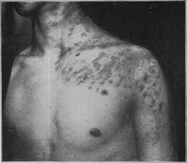
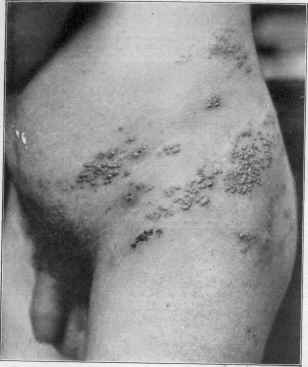
Fig. 84.—Herpes zoster (cervicobrachialis), in a young man aged about twenty-five,
involving neck, shoulders, and upper part of the arms, of eight days’ duration. The
vesicles are small and numerous, some not going beyond the papulovesicular stage, and
closely grouped and coalescent; some hemorrhagic
in zoster of the extremities, especially the upper (Schwimmer, Gibney,
Broadbent, Joffroy, Weiss, and others), and in connection with truncal
zoster; exceptionally hemiplegia, paralysis of bladder, etc, have been
noted (Duncan, Davidsohn).
Etiology.2—Herpes zoster occurs at all ages and in both sexes, but
1 Strübung's paper, “Herpes Zoster und Lähmungen motorischen Nerven,”
Deutsches Archiv für klin. Medicin, 1885, vol. xxxvii, p. 513, refers to most cases pub
lished up to that date; Ebstein, “Zur Lehre von den nervösen Störungen beim Herpes
Zoster mit besonderer Berücksichtigung der dabei auftretenden Facialslähmungen,”
Virchow's Archiv, 1895, vol. cxxxix, p. 505, also gives review of the subject and litera
ture references; Hunt, Jour. Amer. Med. Assoc., 1909, vol. liii, p. 1456, gives a short
preliminary analytic note of 158 collated cases.
2 Clinical analyses bearing upon frequency, etiologic factors, regions involved, etc;
Greenough (255 cases), Boston Med. and Surg. Jour., Oct. 5, 1889—abstract in Jour.
Cutan. Dis., 1889, p. 426; Cantrell (193 cases—observed in services of Duhring, Van
Harlingen, Stelwagon, and Cantrell), Philada. Med. Jour., March 26, 1898. Of the 62
zoster pectoralis cases in Cantrell's analysis, 58 were in males. Max Joseph, ibid.,
1902, vol. x, p. 597; W. Pick, Prag. med. Wochenschr., 1904, p. 219. Among the
HERPES ZOSTER
353
is much more common in males. It is probably most frequent between
the ages of eight and thirty and not at all uncommon after forty; it is
only exceptionally observed in early infantile life. It is not an uncom
mon disease, constituting about 1 to 1.5 per cent, of all skin cases. It
seems much more frequent during spring and late autumn and winter,
and especially during damp, changeable weather. Many causes are
given by different observers for the production of this disease; among
the most important may be mentioned atmospheric changes, exposure
to cold and wet, sudden checking of perspiration, traumatism, peripheral
nerve irritation or injuries (Weir Mitchell, Keen, Picaud, Janin, Bulkley,
Liveing, Köbner, Touton1), pulmonary disease (Leudet, Leroux), in
testinal parasites (Duryee), malaria (Colombini, Winfield2), carbonic
acid gas poisoning (Leudet, Sattler), and the administration of arsenic
(Hutchinson, Dutworth, Gerhardt, Crocker, Zeisler, Nielsen, O‘Donovan,
and many others).3 I have myself met with several instances of its
arsenical production. It may also doubtless arise from reflex irritation,
from functional or organic disease of other organs (Bulkley, Jewell).
In recent years there has been a growing belief that the disease, some
times at least, is of infectious origin, which I believe must be accepted
as probable.
Pathology.—The pathology of this disease has received consid
erable study (Bærensprung, Kaposi, Haight, Robinson, Danielssen,
Weidner, Wyss, and others). The conclusions, in the main, are that
the disease is usually a descending acute neuritis, provoked by various
causes, and that the process has its beginning most frequently in the
ganglionic system—in the cervical or spinal ganglia—finally reaching
the terminal branches with a production of the cutaneous phenomena.
valuable papers on etiology, of recent date, must be mentioned that by W. G. Hay,
Jour. Cutan. Dis., 1898, p. 1 (with good bibliography); Van Harlingen (etiology and
nature), Amer. Jour. Med. Sci., 1902, vol. cxxiii, p. 141; Head, Clifford Allbutt‘s Sys
tem of Medicine, vol. viii; Evans, Brit. Jour. Derm., 1905, p. 198; Knowles, “Herpes
Zoster; A Report of 286 Cases, with a Review of the Unusual Features of the Disease,”
Penna. Med. Jour., May, 1912 (with references); males 205 in 286 cases, 52 cases be
tween ages of twenty to thirty, 3 cases under the age of one, the youngest in a male
aged four months; the most cases (34) occurred in August, the smallest number
(13) in December, and 80 of the cases were observed in three summer months.
1 Weir Mitchell, Injuries of the Nerves and their Consequences, Philadelphia, 1872;
Picaud, Des eruptions cutanêes consécutives aux lesions traumatiques, Paris, 1875.
2 Winfield‘s investigations, New York Med. Jour., 1902, vol. lxxvi, p. 191 (33 cases),
indicate that 40 per cent, of cases show malarial plasmodia in the blood; the literature is
reviewed.
3 Nielsen, “Ueber das Auftreten von Herpes Zoster während Arsenikbehandlung,”
Monatshefte, 1890, vol. xi, p. 302; abbreviated translation in Sydenham Soc‘y‘s Selected
Monographs on Dermatology, London, 1893, p. 167. The writer gives 10 cases of
his own, and references of other cases. The paper is valuable as proving conclu
sively that arsenic can produce zoster; in 557 psoriasis cases taking arsenic, 10 cases of
zoster developed, while in 220 cases otherwise treated zoster was not noted. See also
Rasch, “Contributions a l‘étude des dermatoses d‘origine arsénicale,” Annales, 1893,
p. 150; Méneau, “Dermatoses arsénicales,” ibid., 1897, p. 345; Gerhardt, “Ueber
bläschenförmige, gruppenweise Hautausschläge nach Arsenvergiftung,” Charité-
Annalen, Berlin, 19, Jahrgang, 1894; Sequeira, Brit. Jour. Children's Dis., April, 1904,
records an attack of zoster associated with a generalized bullous eruption, except the
face and extremities, from prolonged administration of arsenic; the zoster was in the
lumbar region corresponding to Head's first lumbar area on the right side. See also
Zeisler‘s paper (“Zoster Arsenicalis,” Jour.Cutan. Dis., 1907, p. 515, with references),
reporting 11 cases.
23
354
INFLAMMATIONS
Investigations (Mackenzie, Head)1 point to a relationship between the
tender areas of visceral disease and the eruptive patches of zoster, the
pain fibers of the skin and viscera being in close connection or associa
tion.2 Clinical observation shows that the eruption does not always fol
low the distribution of one nerve, nor even that of interbranching nerves,
and sometimes the eruption lightly overlaps the median line; this is
doubtless due, as J. Mackenzie‘s investigations,3 and also those of Head
and Campbell,4 show, to some interlocking of nerve-fibers at their origin.
In most cases of zoster the ganglia usually show softening, enlarge
ment, and inflammation, and the nerves are inflamed and thickened.
In the traumatic and also in other instances the ganglia are not involved,
the peripheral nerves alone being the seat of pathologic changes (Charcot,
Weir Mitchell, Pitres and Vaillard, Curschmann and Eisenlohr). It is
probable, I think, that future observations and investigations will show
that many of the zoster-like eruptions, among which are probably to be
placed the recurrent cases, are not examples of true zoster, as already
pointedly suggested by Grindon, Hartzell, Duhring, Hay, and others, but
that if those due to traumatism and other mechanical irritative causes are
eliminated, there will remain the clear-cut typical cases representing a
systemic disease of infectious origin. Numerous examples and clinical
grounds support this view (Rohé, Erb, Jamieson, Landouzy, Barth,
Walther), and it receives further strength from the fact that the disease
occasionally is observed in epidemic form (Neligan, Gauthier, Kaposi,
Weis, Blaschko).5 The fact that zoster occurs but once in a lifetime, the
1 Mackenzie, Med. Chronicle, 1892, vol. xvi, p. 293; Head, Brain, parts i and ii,
1893, vol. xvi, p. 129, and (Herpes Zoster) CliffordAllbutt, System of Medicine.
2 Curtin, “Herpes Zoster and Its Relation to Internal Inflammation and Diseases,
Especially of the Serous Membranes,” Amer. Jour. Med. Sci., 1902, cxciii, p. 264,
reports cases having a clinical bearing on this point; 10 cases associated with various
diseases, as pleuritis, peritonitis, Bright's disease, appendicitis, and esophageal cancer.
In this connection it is interesting to note that Riehl, Münch, med. Wochenschr., 1904,
p. 1105, states that in 481 cases of croupous pneumonia in the Munich Hospital in from
30 to 40 per cent, herpes zoster occurred, generally appearing on the third or fourth
day, and most commonly in the areas supplied with the second and third divisions of
the trigeminus, especially that supplied by the infra-orbital nerve; it had no prognostic
significance; and it is scarcely ever encountered in the pneumonia of children and old
people; see also paper on similar subject by Howard, Amer. Jour. Med. Sci., 1903, vol.
cxxv, p. 256.
3 James Mackenzie, “Herpes Zoster and the Limb Plexuses of Nerves,” Jour, of
Path, and Bacteriol, 1893, vol. i, p. 332.
4 Head and Campbell, The Pathology of Herpes Zoster and Its Bearing on Sensory
Localization, Brain, 1900, vol. xxiii, p. 333 (with illustrations).
6 Some literature bearing upon its infectious and epidemic character: Walther, Allg.
med. Central-Zeitung, 1878, vol. xlvii, p. 394, an observation of 12 to 15 cases (all
students) in three months—no other cases in a period of nine months; in one series
especially reported, a student, after having had an attack, moved from his dwelling;
another later moving in developed the disease; circumstances requiring this student to
leave, the next student taking the same quarters shortly after presented an outbreak.
Kaposi, Wien. med. Wochenshr., 1889, pp. 962 and 1002 (an epidemic of 40 cases);
Weis, Archiv, 1890, vol. xxii, p. 609 (epidemic of 15 cases and some literature refer
ences); Erb, Neurologisches Centralblatt, 1882, vol. i, p. 529 (2 instances in which
mother and daughter developed the disease at about the same time); Pfeiffer, Die Ver-
breitung des Herpes Zoster längs der Hautgebiete der Arteries, Jena, 1889 (based upon 117
cases); also refers to its epidemic and infectious character, and refers to cases; Barth,
Union médicate, 1883, vol. xxxvi, p. 809; Rohé, Arch. Derm., 1877, p. 318; Landouzy,
Semaine médicate, Sept., 1883; Hay, loc. cit.; Wasielewski, Herpes Zoster und dessen
Einreihung unter die Infections Krankheiten, Jena, 1892; Sachs (epidemic in Breslau,
69 cases), Zeitschr. für Heilk., 1904, p. 383.
HERPES ZOSTER
355
usually associated adenopathy, and not infrequently observable sys
temic disturbance, though slight, are, as Hay states, in favor of the infec
tious character of the disease. Exceptionally, it is true, recurrences
have been noted (Kaposi, Behrend, Düring, Nieden, Pernet, Crocker,
Hartzell, Grindon, and others),1 but it is not improbable, as Hartzell
intimates, that many such
cases are of traumatic origin.
It seems, indeed, that any
thing which may bring
about an irritable or in
flamed state of the Gasse-
rian ganglion, spinal gang
lia, nerve-tract, or per
ipheral branches may be
responsible for the erup
tion.
This requisite nerve irri
tation may also be produced
by pressure of tumors
(Eisenlohr and Cursch-
mann). The disease has
also been observed to occur
in myelitis (Hardy, Weid-
ner), hemiplegia (Duncan,
Payne), and in tapes (West-
phal, Bernhardt).
The lesions show (Biesiadecki, Haight, Robinson, Lesser, Kopp,
Lassar, Hartzell, Gilchrist, Unna) some differences from the vesicles
of other diseases. The process begins in the lower rete layer, and
apparently in the papillary layer, but the inflammatory involvement
of this latter is thought to be secondary. The epithelial cells, through
colliquation, undergo enlargement,—ballooning (Unna),—and finally,
1 Grindon, “Recurrent Zoster,” Jour. Cutan. Dis., 1895, pp. 191 and 252, gives an
admirable analysis of recorded cases—61 in all. It shows that most of such cases
cannot be considered as examples of true zoster; a good bibliography is appended.
Vörner, “ Uber wiederauftretenden Herpes Zoster, insbesondere über Zoster ery-
thematosus und Zoster vegetans,” Munch, med. Wochenschr., 1904, p. 1734, reports 3
cases of 3 recurrences in the same region; nervus auricularis magnus; nervus frontalis;
zoster buccalis. In one instance the relapses were of erythematosus patches (zoster
erythematosus recidivus); and in the case of zoster buccalis in one relapse the lesions
were of a vegetating character (zoster vegetans). See also paper, “Du zona récidivant,”
by Hirtz and Salomon, Bull, et mém. soc. d. Hop. de Paris, 1902, 35, vol. xix, p. 206;
and Fabre, “Les recidives du zona,” Bull. Acad. de méd., 3d S., vol. xlix, p. 589, and
Bull, méd., 1903, vol. xvii, p. 376; Einis, “Ueber Herpes Zoster recidivus,” Allg. med.
Centr.-Ztg., 1904, vol. lxxiii, p. 313; Duhring believes (Cutaneous Medicine, part ii, p.
482) that these anomalous, neurotraumatic cases should be classed distinct from
zoster, and suggests the name “dermatitis vesiculosa neurotraumatica,” an example
of which he recently reported (Internat. Med. Mag., March, 1892); Spitzer, “Ein Fall
von recidivirendem Herpes Zoster am Zeigefmger der linken Hand,” Dermatolog.
Centralbl., 1904, vol. viii, p. 74, reports a case in point—there were 5 recurrences in a
year in the district of the musculus radiobrachialis on the index-finger, with, at the
same time, a tenderness of the forearm, with a distinct hyperesthesia of the surface
corresponding to the ramifications of the radial nerve; tactile pain, and thermic senses
much more intense than on the sound side. Grindon (supplementary paper), Jour.
Missouri State Med. Assoc, 1906, No. 8.
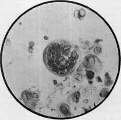
Fig. 85.—Herpes zoster; degenerated epithe
lial, protozoa-like cells found at the sides and base
of vesicle; one resembling a sporocyst (courtesy of
Dr. M. B. Hartzell).
356
INFLAMMATIONS
from pressure and traction, assume various shapes. Some of the degene
rating cells are thinned or flattened out, and small cavities result; soon
these division walls break, and the complete vesicle is produced. The
base of the lesion may be a thin layer of the smaller ballooned epithelial
cells or the papillæ, which latter may project slightly into the cavity;
the roofwall is formed of the corneous layer, to which may be attached
some of the degenerate epithelia. The contents of the lesion consist of
serum, epithelial cells, and later some or many pus-corpuscles, and, in the
hemorrhagic cases, blood-corpuscles. In more especially these latter
cases the upper and sometimes the entire corium undergoes degenerative
and destructive action, and ulceration and consequent scarring result.
Some of the peculiar epithelial cells found resemble protozoa, but their
alleged parasitic character (Pfeiffer) has been disproved, as they have been
shown to be degenerated or altered epithelia (Török, Hartzell, and
Gilchrist).1
Diagnosis.—The usual features of herpes zoster—the frequently
prodromal or accompanying neuralgic pain, the grouped vesicles on
inflammatory patches following the peripheral distribution of one or
two nerves, and exhibiting no tendency to spontaneous rupture, and the
limitation to one side of the body—are quite characteristic and render the
diagnosis a matter of no difficulty. On the face it might be confounded
with an extensive herpes facialis, but in this latter the onesided distri
bution of zoster is usually wanting, likewise the neuralgic pain; the dis
tribution on or about the lips, common in herpes facialis, is infrequent
in zoster. But it must be confessed that occasional cases are encountered
in this region which are somewhat puzzling and which could apparently
be placed under either head.
In those instances in which there may be but two or three patches,
and in which the lesions are small and abortive, scarcely, if at all, reach
ing the vesicular stage, a slight resemblance to papular or vesicopapular
eczema is noted. Eczema, however, rarely consists of distinct or such
sharply defined patches or areas, and is slow in its advent, course, and
disappearance, and the subjective symptom of troublesome itching, al
most invariable in eczema, is usually wanting in zoster. While abortive
zoster is abortive as regards the lesions, it possesses the other features of
the disease, as named above. The beginning symptoms—pain and
neuralgia—of zoster pectoralis have, sometimes been mistaken for incip
ient pleurisy, and such error should, therefore, be guarded against.
Prognosis.—This is almost invariably favorable, the symptoms
usually disappearing in two to four weeks. In extensive cases, and in
those in which new outcroppings present for several days or more,
however, the duration is prolonged to one or two months; and in hemor-
rhagic and ulcerative cases, especially in old people, in whom these types
are commonly seen, while the termination is, as a rule, favorable, a fatal
ending through exhaustion or septic conditions is possible. In zoster
involving the eye the outlook is not always certain, as uselessness or
1 Pfeiffer, Monatshefte, 1887, vol. vi, p. 589; Török (quoted in Brooke‘s Hamburg
letter), Brit. Jour. Derm., 1890, p. 120; Hartzell, Jour. Cutan. Dis., 1894, p. 369;
Gilchrist, Johns Hopkins Hospital Reports, 1896, vol. i, p. 365.
HERPES ZOSTER
357
destruction of this organ may ensue, and exceptionally septic infection,
meningitis, and death. The possibility of persistent neuralgia or other
sensory and rarely motor disturbances following the eruption is to be
kept in view. It is to be said, however, that in a large number of the
cases observed the disease is benign, and the patients go about and suffer
but little inconvenience.
Treatment.—The mild cases rarely require any constitutional
treatment. In those more severe systemic remedies should always
be prescribed, the character of the treatment depending, for the most
part, upon the indications presented by the individual patient. The
chief remedies prescribed, independently of general principles, are those
directed toward invigorating the nervous system. Zinc phosphid, 1/5
grain (0.013) (Thomsom, Bulkley) every three or four hours, seems
at times to be of service. In other cases large doses of quinin and
strychnin will be found to be useful; arsenic is also thought to be of
service. Zinc phosphid and quinin prescribed together has seemed to
me beneficial and a good routine practice. In cases in which pain is
an urgent symptom, it may be necessary to prescribe potassium bro-
mid, chloral, sulfonal, and even rnorphin; in extreme instances of this
character the hypodermic administration of the last-named drug will
be demanded. Antipyrin, phenacetin, and acetanilid may also be used
for this purpose, and these several drugs, it has been alleged, not only
relieve the pain, but may favorably influence the disease. Jarisch
speaks well of the conjoint administration of 7 or 8 grains (0.465-0.53)
each of antipyrin and sodium salicylate, three or four times daily. Lassar
commends highly full doses of sodium salicylate.
External applications are of importance in all except the extremely
mild and abortive cases; these latter usually require but little, if any,
treatment. The lesions rarely need to be opened. As a rule, the sole
object in view in the use of local applications is protection to the parts.
This may be accomplished in mild or average cases by the free use of
a dusting-powder of equal parts of zinc oxid, boric acid, and talc, over
which may be placed a layer of cotton thoroughly drenched with the
same powder; this is kept in place by a gauze bandage. This is to be
changed daily or every few days, without disturbing the parts unless
soiled or offensive, in which event washing with saturated solution of
boric acid is advisable; this is, however, rarely required. In mild cases
but one or two renewals of the dressings are necessary. A wet dressing
of carbolized alcohol (Leloir), 0.5 to 1 per cent, strength, or one to several
grains of menthol to the ounce of alcohol, may be used; this is applied
on compresses and covered with gutta-percha tissue, and renewed
several times daily. In other cases ointments seem to give the most com
fort, such as zinc oxid ointments, with 1 or 2 drams (4.-8.) of starch to
the ounce (32.), and to which also in painful cases may be added from 5
to 20 grains (0.32-1.3) of opium, or from 3 to 10 grains (0.2-0.66) of
menthol to each ounce (32.). Such a dressing need be changed but once
or, at most, twice daily. Fabre commends, for allaying the pain, painting
over the areas a mixed solution of 1 per cent, adrenalin and 2½ per cent,
cocain. A valuable method of treatment consists in the application,
358
INFLAMMATIONS
five to ten minutes daily, or twice daily, of a mild galvanic (constant)
current, 1 to 3 milliampères, the positive electrode being placed as near
as possible to the main nerve-supply of the part, and the negative being
gently move to and fro over the diseased area; it favorably influences the
pain and seems to modify the course of the disease. For the pain that
sometimes follows in the wake of the disease the galvanic current also
often gives prompt relief.
But first, if you want to come back to this web site again, just add it to your bookmarks or favorites now! Then you'll find it easy!
Also, please consider sharing our helpful website with your online friends.
BELOW ARE OUR OTHER HEALTH WEB SITES: |
Copyright © 2000-present Donald Urquhart. All Rights Reserved. All universal rights reserved. Designated trademarks and brands are the property of their respective owners. Use of this Web site constitutes acceptance of our legal disclaimer. | Contact Us | Privacy Policy | About Us |