| MEDICAL INTRO |
| BOOKS ON OLD MEDICAL TREATMENTS AND REMEDIES |
THE PRACTICAL |
ALCOHOL AND THE HUMAN BODY In fact alcohol was known to be a poison, and considered quite dangerous. Something modern medicine now agrees with. This was known circa 1907. A very impressive scientific book on the subject. |
DISEASES OF THE SKIN is a massive book on skin diseases from 1914. Don't be feint hearted though, it's loaded with photos that I found disturbing. |
POROKERATOSIS
Synonyms.—Hyperkeratosis eccentrica; Keratodermia eccentrica; Hyperkeratosis
figurata centrifuga atrophicans; Fr., Porokératose; Hyperkeratose figureé centrifuge
atrophiante.
Under this title Mibelli4 called attention to a rare and undescribed
variety of hyperkeratosis, presenting in small, eccentrically developing
areas. Simultaneously appeared also a description of the malady by
Respighi,5 and since these first cases new reports have been made by
the same observers and by Hutchins,6 Reisner,7 Joseph,8 Gilchrist,9
1 Andeer, “Resorcin bei Ichthyosis,” Monatshefte, 1884, vol. iii, p. 365.
2 Jamieson, Diseases of the Skin.
3 Unna, “Aphorismen über Schwefeltherapie und Schwefelpräparate,” Monatshefte,
1883, vol. ii, p. 197.
4 Mibelli, Giorn. ital., 1893, p. 313; Monatshefte, Nov. 1, 1893; International Atlas
of Rare Skin Diseases, 1893, vol. ix, plate xxvii; Annales, 1905, p. 503.
5 Respighi, Giorn. ital., 1893, p. 356, and 1895, P- 69, and Monatshefte (translation
of first paper), 1894, vol. xviii, p. 70 (with case illustration and histologic cuts).
6 Hutchins, Jour. Cutan. Dis., 1896, p. 373 (with case illustration and a review of
the published cases, with references).
7 Reisner, Ein Fall von Porokeratosis, Inaug. Dissertation, Strassburg, 1896.
8M. Joseph, Archiv, 1897, vol. xxxix, p. 335 (case illustration and 11 histologic
cuts; review of other published cases and references).
9 Gilchrist (preliminary paper), Bull. Johns Hopkins Hosp., 1897, p. 107; (main
paper) Jour. Cutan. Dis., 1899, p. 149 (with case illustrations; 11 cases in one family
(four generations); 5 histologic cuts and bibliography).
572
HYPERTROPHIES
G. W. Wende,1 and Rasch.2 The disease is extremely slow and insidious,
appearing first as a trifling, superficial but slightly elevated, warty-
looking formation, or as thin, callous spots. These gradually enlarge,
sometimes months or years elapsing before reaching conspicuous dimen
sions. The spots extend by a peripheral thickened “seam,” “dike,”
or “wall,” and, usually leave an atrophic, generally slightly or moderately
calloused, center. In some cases the inclosed portion consists of some
what atrophic glossy epidermis, in others a trifle thickened, but per
ceptibly depressed, and sometimes presenting a dotted appearance.
The border is rather sharply defined against the outlying sound skin,
and is hard or horny in character, with often a linear horny ridge, in the
middle line of which there is a narrow sulcus, and in this very often a
thin, horny, thread-like or cord-like elevation, somewhat irregularly di-
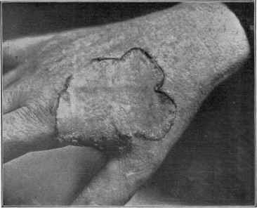
Fig. 137.—Porokeratosis (courtesy of Dr. G. W. Wende).
viding the sulcus into two lateral halves. In this thus inclosed line are
often found here and there round, millet-seed or smaller sized blackish
epidermic concretions, which can be picked out (Hutchins). Occasion
ally, too, these or similar concretions or minute wart- or papillary-like
corneous projections are found imbedded in the inclosing horny lateral
elevations of the seam or border, as well as within the atrophic hardened
inclosed portion. Sometimes the border is distinctly wall-like, its in
closing side rather sharply perpendicular, and the other side rapidly,
but not precipitously, merging into the surrounding skin. In contour
1 G. W. Wende, Jour. Cutan. Dis., 1898, p. 505 (with case illustration and histologic
cut and bibliography).
2 Rasch, Pester med.-chirurg. Presse, 1898, p. 626—abs. in Jour. Cutan. Dis., 1898,
p. 547; Heidingsfeld, Jour. Cutan. Dis., Jan., 1905, reviews the subject and gives com
plete bibliography; Brocq and Pautrier, Tribune Médicate, June 22, 1907, case, young
woman, patches on face, nucha, left hand, and forearm (early references).
POROKERATOSIS
573
the areas are sometimes fairly well rounded, but often somewhat wavy,
others of irregular squarish shape, and others still more irregular in out
line. The inclosed portion almost always shows slight or moderate
epidermic thickening and variable atrophy, with slight scaliness or fairly
smooth; but occasionally it is but little changed, having a faintly atrophic
appearance, and the hairs may or may not have disappeared. The
color of this part may be grayish white, dirty gray, sometimes with a
brownish hue, and exceptionally, more especially in ill-developed spots,
a pinkish tinge. The “seam,” “wall,” or “dike” may be dirty gray or
brownish gray, and is usually quite pronounced, horny, and elevated;
in others—ill-defined spots—it may appear simply as a loose rim of
epidermis, made up of one or several layers, and the free edges directed
inward and slightly upward. In one and the only instance under my
own care there was but a single patch, of years’ duration, and seated
on the dorsal surface of the hand, between the metacarpal bones of the
thumb and forefinger; the patch, about an inch to an inch and a half in
diameter, was irregularly rounded, with a pronounced wavy elevated
border, with an ill-developed irregular and broken sulcus, more or less
studded with hard concretions or seed-wart-like epidermic accumula
tions. The inclosed portion was depressed, somewhat horny, slightly
scaly, uneven, and with here and there the imbedded minute, warty-
looking concretions just referred to. The broken character of the dike
or wall is not unusual, although it is often continuous.
The favorite regions are the hands and feet, more especially the
dorsal aspects, but also not uncommonly on the palmar and plantar
surface as well. The patches occur on other parts, however, as the face,
limbs, and trunk. But one or several may be present, or there may
be many in various sizes and of somewhat general distribution. As a
rule, they do not exceed one to several inches in diameter, and sometimes
remain much smaller. There is usually slight, continuous extension, but
sometimes, after reaching a certain development, they remain practically
stationary; where several contiguous patches coalesce a considerable area
may result. As a rule, there are no subjective symptoms, although in
some cases variable itching has been noted. The sweat and sebaceous
secretions of the affected areas are more or less in abeyance. While
the integument alone is usually the seat of the malady, Respighi and
Ducrey1 have shown that it is not at all uncommon for lesions to be
seen on the mucous membranes of the mouth also; in 3 of the 4 cases
observed by them, the lesions occurring here appearing as opalescent,
rounded, or irregularly rounded patches, each inclosed by a distinct
white raised line or border, sometimes interrupted, and surrounding
which there is usually a slight zone of hyperemia. Its course here, as
on the skin, is slow and chronic, and apparently gives rise to no incon
venience. Mibelli2 also found, in an extensive case, lesions in the mouth
as well as on the glans penis.
1 Respighi and Ducrey, Annales, 1898, pp. 1, 609, and 734—an exhaustive account
of the disease,—clinical and histologic,—3 case illustrations, and 48 histologic cuts.
2 Mibelli, Archiv, 1899, vol. xlvii, pp. 1 and 231; review of the disease, 5 case illus
trations, and 6 histologic cuts.
574 HYPERTROPHIES
Etiology and Pathology.—But little is known as to the cause
of the disease, although a hereditary tendency was indicated in Res-
pighi‘s case, as the father of the patient had similar lesions; the hered
itary factor has been convincingly shown by Gilchrist in his report of
ii cases in one family—in 4 generations. Respighi and Ducrey also
report an instance of the malady occurring in several generations. It is
a rare disease, the cases of Hutchins, Gilchrist, Wende, and my own1
apparently being all that have been recorded in this country. It is
seemingly not so rare in Italy, but is scarcely known in England, France,
and Germany. It is met with in both sexes and at all ages, but it has
its beginning more frequently in early life. There is no direct evidence to
prove that the disease is parasitic, although in 30 experimental inocula
tions made by Wende on 4 different individuals one proved successful,’
but as this was on the affected patient, it is not wholly conclusive; it is
possible that the local irritation produced may have been sufficiently
potential in a predisposed skin, although it is true in this instance the
inoculation, made on the unaffected hand, was positive only after 10
unsuccessful attempts. Respighi‘s experiments in transplantation were
without result. Examinations made for micro-organisms have been
uniformly negative. The predominance of parts subject to pressure
and friction as sites of the eruption, as the hands and feet, appears to
indicate that these factors may be contributory.
Histologic examinations were made by almost all the observers
named, and their conclusions in the main agree that the malady is a
special form of hyperkeratosis, and affecting chiefly the lower horny
and upper rete layers, although all parts of the epidermis, especially
about the sweat-gland ducts, which are often plugged up with horny
epithelium, share in the process. The hair-follicles and sebaceous
glands also show involvement. The papillary layer of the derma is
almost obliterated in the central area (Respighi). Tommasoli2 was
inclined to question the individuality of the affection and the identity
of the various cases reported, believing that they were unusual exam
ples of other keratoses, such as ichthyosis, linear nævus, etc., but the
clinical features, as well as the histologic findings, and the behavior
and course of the disease, as Mibelli3 convincingly showed, are strikingly
different from any other known malady.
Prognosis and Treatment.—The malady, as will have been
seen, is a persistent one, with but little, if any, tendency to spontaneous
disappearance, but beyond the disfigurement which it causes need give
rise to no anxiety. Occasionally some of the efflorescences may dis-
11 saw this case in 1887 or 1888, on two occasions only, but was puzzled by it and
did not recognize its nature, intending to publish it later; the papers of Mibelli and
Respighi appearing subsequently showed me that I had missed an opportunity of pri
ority. Recently (1909) Dr. H. H. Rutherford, U. S. A., was kind enough to send me a
photograph of a case in a young soldier aged twenty-six, showing an elongated area
at base of thumb, extending on the dorsal surface of hand; it began at the age of sixteen
as a small, warty pimple, which gradually scaled off and became callous, with a thick
ened seam bordering it.
2 Tommasoli, Comment, clin. d. mal. cut. e. gen. ur., 1894, ii, No. 1.
3 Mibelli, “Ueber die Porokeratose (Antwort auf eine Kritik),” Monatshefte, 1895,
vol. xx, p. 309 (with references).
ANGIOKERATOMA
575
appear (Joseph). The lesions on the palms and soles are sometimes
rendered painful by the constant pressure, friction, etc
The treatment of the affection is somewhat uncertain as to result
unless surgical measures are employed. Joseph was not able to obtain
any result from salicylic acid and other keratolytics, but with excision
there was no recurrence. Gilchrist found that the lesions always re
turned after thorough curetting and subsequent application of silver
nitrate, a plan which was tried first. Excision proved satisfactory, but
naturally left scars. In 2 of his cases the electric needle was used with
excellent results, causing very little scarring, and there was no return.
But first, if you want to come back to this web site again, just add it to your bookmarks or favorites now! Then you'll find it easy!
Also, please consider sharing our helpful website with your online friends.
BELOW ARE OUR OTHER HEALTH WEB SITES: |
Copyright © 2000-present Donald Urquhart. All Rights Reserved. All universal rights reserved. Designated trademarks and brands are the property of their respective owners. Use of this Web site constitutes acceptance of our legal disclaimer. | Contact Us | Privacy Policy | About Us |