| MEDICAL INTRO |
| BOOKS ON OLD MEDICAL TREATMENTS AND REMEDIES |
THE PRACTICAL |
ALCOHOL AND THE HUMAN BODY In fact alcohol was known to be a poison, and considered quite dangerous. Something modern medicine now agrees with. This was known circa 1907. A very impressive scientific book on the subject. |
DISEASES OF THE SKIN is a massive book on skin diseases from 1914. Don't be feint hearted though, it's loaded with photos that I found disturbing. |
SPOROTRICHOSIS
It is due to Schenck’s1 (1898) paper, the Brayton2 (1899), Hektoen
and Perkins (1900),3 and De Beurmann and Ramond (1903)4 reports, and
more recently to the distinguished work of De Beurmann and Gougerot,5
1 Schenck, “On Refractory Subcutaneous Abscesses Caused by a Fungus, Possibly
Related to Sporotricha,” Johns Hopkins Hosp. Bull., 1898, p. 286.
2 Brayton, “Chronic Abscesses,” Indianapolis Med. Jour., 1899, vol. xviii, p. 272.
3 Hektoen and Perkins, “Refractory Subcutaneous Abscesses Caused by Sporothrix
Schenckii: A New Pathogenic Fungus,” Jour. Exper. Med., 1900, p. 77.
4 De Beurmann and Ramond, “Absces sous-cutanés multiple d’origine mycosique,”
Annales, 1903, p. 678.
5 De Beurmann and Gougerot, Bull, de la Soc. Franc, de Derm., Jan. 3, 1907, p. 26
(dermic sporotrichoses); Annales, 1906, pp. 837, 914, and 933 (subcutaneous sporotri-
choses; complete exposition with many illustrations); ibid., 1907, pp. 497, 603, and 654
(“sporotrichoses tuberculoides,” case and histologic illustrations, review and references);
Tribune Medicale, Nov. 2, 1907, p. 693 (etiology and pathogeny); Bull, de la Soc. Med.
des Hôp. de Paris, June 7, 1907, p. 585 (mucous membranes); De Beurmann, Gougerot,
and Vaucher, ibid., Oct. 25, p. 107 (in cat); ibid., May 22, 1908, p. 718 (in rat); ibid.,
June 5, p. 800 (in rats—experimental); De Beurmann and Gougerot, ibid., May 28,
1908, p. 733 (wide diffusion; North and South America); Annales, 1909, p. 81 (acute,
subacute, and sluggish types, and comparison with other somewhat similar infections;
review and references); Archiv, 1911, cx, p. 25 (general review); “Les Sporotrichoses,”
Paris, Libraire Felix Alcan, 1912; and many other papers (chiefly case reports).
1168
PARASITIC AFFECTIONS
Monier-Vinard,1 and others,2 that the importance of this malady and its
pathogenic fungus, the sporotrichium, is being gradually fully recognized.
Symptoms.—While the symptomatology may vary considerably
within certain limits, there is a family likeness: quite often a case pre
sents an ill-defined symptom medley of a tuberculous and a syphilitic
gumma, tuberculosis verrucosa cutis, and a variable degree of pyo-
genic inflammation. In one set of cases the malady takes its origin
1 Lesne and Monier-Vinard, Bull, de la Soc. Anat., May, 1906, p. 422 (multiple
chronic abscesses); Bull, de la Soc. Med. des Hôp. de Paris, March 21,1907, p. 268
(subcutaneous); Gaudier and Monier-Vinard, ibid., May 2, 1907, p. 353 (2 cases;
cutaneous and visceral sporotrichosis; one had pulmonary tuberculosis); Monier-
Vinard, Presse Mêdicale, July 6, 1907, p. 426 (clinical types and diagnosis); Rubens-
Duval and Monier-Vinard, Bull, de la Soc. Méd. des Hôp., Oct. 25, 1907, p. 1074; and
several others.
2Adamson, Brit. Jour. Derm., 1908, p. 296, gives an excellent résumé, with full
bibliography; Mewborn, Jour. Cutan. Dis., 1908, p. 140, also a good, but somewhat
briefer, one with partial bibliography; Chipman, “A Résumé of the Views of De Beur-
mann and Gougerot on the Subject of Sporotrichosis,” Jour. Cutan. Dis., 1912, p. 339.
Among other case reports and valuable contributions during the past few years are:
Arndt, Berlin, klin. Wochenschr., 1909, No. 44 (preliminary report of first German
case, patient male, aged twenty-nine, with gummatous type on right arm); and later
report, “Beiträge Zur. Kenntniss der Sporotrichose der Haut mit besonderer Berück-
sichtigung der Lymphangitis Sporotrichotica; expermentelle Sporotrichose,” Derma-
tolog. Zeitschr., 1910, H. 1 and 3 (histologic and clinical study with review and résumé
of literature and bibliography; experimental inoculation successful in rats, apes, but
negative in guinea-pigs); and Archiv, 1911, cx, p. 25 (present status of the question and
general review); Kren and Schramek, Wlen. klin. Wochenschr., 1909, xxii, p. 1519 (leg
first, later other parts; nodular and furunculoid); Burlew, S., Cal. Pract., Jan., 1909, p.
1 (abscess on left cheek, and numerous small abscesses on the right anterior leg some
what deep); Trimble and Shaw, Kansas Med. Jour., Sept., 1909; Sutton, Jour. Amer.
Med. Assoc, Sept. 17, 1910, p. 1000 (illustrations; woman aged thirty, beginning on
ball of right thumb, possibly from a splinter); “Sporotrichosis in America,” Jour. Amer.
Med. Assoc, Dec 24, 1910 (2 new cases, with illustrations); and “Sporotrichosis in
Man and in the Horse,” Boston Med. and Surg. Jour., Feb. 9, 1911 (also reports a case in
which infection was probably from a horse); Hyde and Davis, “Sporotrichosis in Man,
with Incidental Consideration of its Relation to Mycotic Lymphangitis in Horses,” Jour.
Cutan. Dis., 1910, p. 321 (with many illustrations, review, and full bibliography; 1 case,
beginning on hand thought to have been derived from a similar infection in a horse);
Adamson, June 1911, p. 182 (case demonstration); and ibid., p. 239 (full report of same
case, Englishman infected in Brazil, beginning in slight wound in back of his right
thumb, with small chain of arm lesions; illustrations of case and fungus); Brit. Jour.
Derm., 1913, p. 33 (case demonstration; second case; disseminated, ulcerative, gumma-
tous type with acute synovitis, woman aged sixty; under potassium iodid case much
improved, but this drug seemed to start up the first trouble); and “A Case of Sporo-
trichosis Simulating Blastomycosis,” Brit. Jour. Derm., 1913, p. 60 (third case; patient
in the United States when first symptoms appeared; first lesions upon right leg followed
by others on trunk and hands; lesions papillomatous); Ofenheim, Lancet, March 11,
1911 (doubtful case); Norman Walker and Ritchie, Brit. Med. Jour., July 1, 1911 (male;
injury of hand, extended up arm); Hodare and Bey, Archiv, 1911, cx, p. 387 (a case
of one and one-half years duration with septicemic symptoms, the eruption being
generalized and consisted of acneiform papules and nodules, many of which were vesico-
pustular, others necrotic and covered with a crust which on healing left small cica
trices and persistent pigmentation, finally recovered); and Dermatolog. Wochenschr.,
Jan. 13, 1912, p. 50—abs. in Jour. Cutan. Dis., April, 1912 (3 cases in same family,
two sisters and a brother, one sister had purplish, lentil-sized nodules on the face for
three months’ duration, some of the lesions being crusted; the other sister had a chest
nut-sized tumor on the ala nasi resembling lupus verrucosus, with smaller lesions on the
face, the hands, and left elbow, and of eight months’ duration; the brother had a single
lesion on the wrist of eight months’ duration; fungus found and cultured); Hamburger,
Jour. Amer. Med. Ascoc, Nov. 2, 1912, p. 1590 (case male, farmer, aged twenty-eight;
chain of lesions on leg similar to arm cases; interesting and valuable summary and ana
lytic tabulation of the American cases, with bibliography); Kenneth Taylor, “Sporo-
trichium Schenkii,” ibid., April 12, 1913, p. 1142 (chiefly on morphology and cultural
characteristics, and review, with references)—a helpful paper to those interested.
SPOROTRICHOSIS 1169
at some point of trifling injury—for example, on the hand.1 The seat
or injury may or may not become sluggishly inflamed, and develop into
a small dermic, sometimes subdermic nodule, and which may or may not
soften or break down; or the point of injury may develop into a sluggishly
inflamed discharging sore; or there may simply result an insignificant
abrasion or sore, which has almost or entirely disappeared before the other
formations have developed. At about the same time or a little later
a subcutaneous nodule will be felt at the lower end of the forearm; this
gradually enlarges to the size of a cherry, flattens somewhat, and is not
much elevated, but may spread out peripherally; it softens, the overlying
skin thinning and becoming of a purplish-red color, and finally breaking
through, the discharge being of a viscid, gelatinous, seropurulent charac-
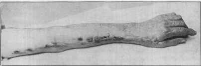
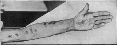
Fig. 311.—Sporotrichosis (Stelwagon-Stout Jefferson Hospital case),
ter. Its formation is successively followed by several or more such for
mations higher up the arm, along the lymphatic vessel, which can
usually be felt as a hard cord. They all may not open, one, two, or
more remaining as cold sluggish subcutaneous tumors, from the size
of a cherry to that of a mandarin orange; the smaller ones usually hard,
the larger ones being of the nature of a partly or more completely softened
indolent abscess. As a rule, however, softening is followed by gradual
1 In Schenck’s case, as well as in the Hektoen and Perkins case, it was a slight injury
to the finger from a nail; and in Brayton’s case it was the puncture of the skin of the
finger by a wire. In an instance at my service at the Jefferson Medical College Hospital
it originated in an injury of the palm, just at the base of the thumb (this case was shown
before the Philadelphia Dermatological Society, Nov. 24, 1908, and appears in Society
Transactions, “Case for Diagnosis,” Jour. Cutan. Dis., 1909, p. 131).
74
1170 PARASITIC AFFECTIONS
extension to the surface, breaking through with a resulting irregular
crateriform opening. It takes generally several weeks to a few months
for the malady to reach this stage. After discharging, a lesion may grad
ually undergo partial involution, and then remain stationary; or it may
extend laterally somewhat as a slightly nodular purplish-red infiltration,
in a measure similar to that seen associated with a broken-down tubercu
lous gland.
In the other cases, by far the larger number, the port of entry—
point of infection—is not discoverable. In one class of these cases,
chiefly described by French observers, the development of the lesions
is about similar to that described, but they are irregularly disseminated
over the limbs and trunk, beginning primarily in the subcutaneous tissue
as somewhat hard, painless nodules, slowly increasing in size; usually
breaking down, the contents discharging through the fistulous opening
as a grayish-yellow homogeneous pus. These cases represent the so-
called “syphiloid” type of De Beurmann and Gougerot. It is not im
possible that exceptionally there may be not more than one such forma
tion, and seated at almost any region; but, as a rule, there are from five
to thirty. Sometimes a number of these may not develop beyond the
size of a large pea to small cherry-sized hard nodules, which can be felt
as an irregular, subcutaneous, nodular chain along an enlarged lymphatic.
Some of the broken-down lesions finally heal, leaving a scar; or they tend
to become papillomatous, resembling tuberculosis verrucosa cutis. In
some instances this fungating tendency is so marked as to give the malady
a distinctly tuberculous aspect, the “tuberculoid” type of De Beurmann
and Gougerot; although in this type the lesions usually have their origin
closer to the surface. In fact, some of the tuberculosis verrucosa type
of lesions may apparently begin as such, or as a small ulcer, resulting
from a rapid breaking down of the surface of a superficial nodule or papu-
lotubercular infiltration. It is more especially this verrucous or papil-
lomatous type that presents some resemblance, usually slight, to blasto-
mycosis.
In another class of cases the lesions develop primitively in the
dermis, as in the Monier-Vinard case. In this instance it presented
as a number of distinctly elevated, moderately sized, roundish nodules
on the face; the surface of the lesions was at first smooth and moist,
but later, after weeks or a few months, rough and irregular with a yel
lowish coating; the scars that followed resembled those left by lupus
tubercles. Another, secondary form—the epidermic form—is also ex
ceptionally observed, in which an irregular ring of vesicopapules or opal
ine vesicles develop around the central opening of the discharging or
discharged nodule, presenting a rough resemblance of trichophytosis.
In rare instances intramuscular abscesses and gumma-like formations
beneath the periosteum (usually a tibial periostitis) have also been noted;
the latter, with one exception only, has been associated with the more
usual subcutaneous abscesses. Exceptionally, a large abscess type (Dor)
has been observed.
Mucous membrane (buccopharynx) lesions have also occasionally
been noted (De Beurmann and Gougerot, Brodier and Gaston, and
SPOROTRICHOSIS 1171
Letulle); De Beurmann and Gougerot discovered (1 case) that the organ
ism may exist in this region in sporotrichosis without producing lesions.
The sporotrichium was found by Monier-Vinard in the expectoration of
a patient (sporotrichosis case) affected with pulmonary tuberculosis,
possibly a mixed infection; it has likewise been found in the sputum of a
sporotrichosis patient when the lungs were apparently healthy. In
a few other instances there seemed to be evidences of systemic and pos
sibly visceral involvement (Massery, Doury, Monier-Vinard, and
Widal and Weill). While the lymphatic vessels concerned may be, and
usually are, affected—a decided lymphangitis sometimes—the glands
are rarely enlarged.
The course of the malady, as is to be already inferred, is slow; and
it is persistent, with only rarely, except as to individual lesions, any
tendency to complete spontaneous recovery.
Etiology and Pathology.—The malady is most frequently seen
in France; the United States (Schenck, Brayton, Hektoen and Perkins,
Burlew, Trimble and Shaw, Hyde and Davis, Pusey, Stelwagon and
Stout, Sutton, and several others) coming next in number of cases;
England (Adamson, Norman Walker) and Germany (Kren and Schramek
and Arndt) so far recording but few cases; the disease is, however, doubt
less worldwide. It is due to a fungus, the sporotrichium, as primarily
discovered by Schenck, and later confirmed by Hektoen and Perkins,
and since thoroughly established by the observations and experimental
investigations of De Beurmann and Gougerot. At first it was generally
accepted that the organism was identical in all the cases, but De Beur-
mann and Gougerot and several others now believe that there are
several forms of the fungus, three of which are thought to have been
identified: sporotrichium Beurmanni in the European and Brazilian
cases, sporotrichium Schencki in the North American cases, and sporo-
trichium indicum in Ceylon cases. It can rarely be demonstrated in the
lesions, but is readily cultured and on almost any of the usual media.1
The parasite has been isolated from the blood of a patient with cutaneous
sporotrichosis (Widal and André). The malady has been produced (De
Beurmann and Gougerot and others) experimentally by cutaneous or
intraperitoneal inoculation in animals (guinea-pig, cat, rat, mouse, and
monkey), the rat being especially susceptible, and the guinea-pig the
least; although De Beurmann has produced a generalized subcutaneous,
gummatous sporotrichosis in a newborn guinea-pig by feeding it upon
milk containing the parasite. In the rat visceral reactions are quite
common. Spontaneous sporotrichosis has been noted in the mule
1 Sabouraud’s peptone-glucose-agar is the best. Streak cultures are made upon
this medium of the pus taken from the suspected lesions, taking the usual precautions
to avoid contamination and avoiding the use of rubber caps over the cotton plugs.
The fungus is best grown at room temperature; small white colonies appear on the
fourth to the eighth day along the line of the streak. They slowly increase in size, and
become convoluted and brown in color. In bouillon the growth may form a veil or a
flocculent down without the medium becoming cloudy. In the hanging-drop the para
site appears as a fine ramified mycelium, with partitions at long intervals. Films from
cultures show long filaments 2 µ broad, together with numerous ovoid spores 5 to 6 µ
in length by 3 to 4 µ broad; here and there single spores, or bunches of three to thirty,
are seen attached bouquet-like to the mycelial filaments by a short pedicle.
1172 PARASITIC AFFECTIONS
(Fontegnot and Carongean), in the dog (Gougerot and Caraven), and
in the rat (Lutz and Splendore); and the saprophytic nature of the fungus
has been demonstrated by its culture upon various animal structures,
such as caterpillars, flies, larvæ, etc., and upon vegetables as well. Hyde
and Davis’ investigations show that some of the American cases of my-
cotic lymphangitis, or epizoötic lymphangitis, in horses are due to this
same Sporotrichium, and are in reality cases of sporotrichosis, thus
furnishing another source for the infection in man. It can be readily seen,
therefore, how easily infection, under favoring circumstances, might
take place, and in many ways either externally or internally. The port
of entry has remained unknown, however, in many cases. It is seen in
both sexes and at almost all ages, although uncommon in childhood; in
74 cases (collected by Sutton) the youngest patient was aged five; the
oldest aged seventy-eight; there were 14 females and 60 males; in 30
instances the initial lesion was on the hand or forearm, and in 11 on the
foot, legs, or thigh. The malady is probably not so rare as recorded ob
servations would indicate, as cases simulative of syphilitic gummata may
have been treated as such with the iodids with recovery, and the error
in diagnosis remain undiscovered.
In their histologic study De Beurmann and Gougerot found that
the malady is of the nature of a chronic, nodular, suppurative affection,
and that the histologic picture showed resemblance, as the affection
often does clinically, to that of tuberculous and of syphilitic lesions.
There seems to be a mixture of the three types of reaction—(1) a lympho-
connective tissue or syphiloid; (2) an epithelioid (with giant-cells) or
tuberculoid; and (3) a polynuclear or suppurative.
Diagnosis.—The peculiar conditions in the hand and arm cases,
already described, are more or less suggestive. In the more general
cases the varying characters of the lesions, due to their different stages,
the cold sluggish nature of the softening and abscess-formation, the cra-
teriform aspect, the slow course, and the usually undisturbed general
health, are to be taken into consideration. The histologic picture is
not distinctive enough to be of much aid. The suspicion aroused, the
diagnosis can be confirmed or disproved by cultural methods.1 The
diseases with which it might most likely be confused are those previously
mentioned: tuberculous and syphilitic affections, simple indolent,
staphylococcic abscesses, and blastomycosis.2 It is to be remembered
that it is not impossible that sporotrichosis may coincidentally also be
present in a subject with syphilis, tuberculosis, or any other disease.
Prognosis and Treatment.—The disease usually responds
1 De Beurmann and Gougerot have found that an early orchitis (in fifteen to twenty
days) in the rat after intraperitoneal inoculation with material from the suspected case
is diagnostic
2 De Beurmann and Gougerot, “Eine Neue Mykose Die Hemisporose,” Archiv,
April, 1910, ci, p. 298,—abs. in Brit. Jour. Derm., 1910, p. 297; under the name “hemi-
sporosis” these observers describe 3 cases due to the hemispora stellata, and which
might possibly be mistaken for the gummatous types of sporotrichosis; the first case
(Gougerot and Caraven) simulated a syphilitic ostitis of the tibia, the second case simu
lated a tuberculous abscess on the neck, and the third case simulated a gumma of the
penis; histologically, there was but little difference in the findings from those of tubercle
or syphilis.
PEDICULOSIS
1173
more or less rapidly to potassium iodid, in medium to large dosage.
Applications externally to the broken-down lesions and ulcers of com
presses of weak lotions of the same drug, or diluted Lugol’s or other weak
iodin solution, are helpful. The x-ray would doubtless be of some aid.
Surgical measures are not to be commended unless the pus-collection
should be large.
But first, if you want to come back to this web site again, just add it to your bookmarks or favorites now! Then you'll find it easy!
Also, please consider sharing our helpful website with your online friends.
BELOW ARE OUR OTHER HEALTH WEB SITES: |
Copyright © 2000-present Donald Urquhart. All Rights Reserved. All universal rights reserved. Designated trademarks and brands are the property of their respective owners. Use of this Web site constitutes acceptance of our legal disclaimer. | Contact Us | Privacy Policy | About Us |