| MEDICAL INTRO |
| BOOKS ON OLD MEDICAL TREATMENTS AND REMEDIES |
THE PRACTICAL |
ALCOHOL AND THE HUMAN BODY In fact alcohol was known to be a poison, and considered quite dangerous. Something modern medicine now agrees with. This was known circa 1907. A very impressive scientific book on the subject. |
DISEASES OF THE SKIN is a massive book on skin diseases from 1914. Don't be feint hearted though, it's loaded with photos that I found disturbing. |
ATROPHIA UNGUIUM1
Synonyms.—Atrophy of the nails; Onychatrophia.
Symptoms.—In atrophy of the nails these appendages may show
various conditions; they may be soft, thin, and brittle, splitting easily,
opaque and lusterless, and sometimes with a worm-eaten appearance.
But one of these characters may be present, or several or all may be
exhibited. It may be congenital or acquired.
Congenital cases are rare, and in such instances it is not uncommon
to find imperfect growth or absence of the hair, and also defective forma
tion of the phalanges. In some of these instances the nails are entirely
wanting, as in Eichhorst’s2 case, although the nail-bed and fold were well
developed, hair and teeth normal, and no hereditary history. In the
congenital hypertrophic cases3 recorded by Nicolle and Halipré and C. J.
White, especially in the series reported by the latter, in addition to hy
pertrophy, atrophic changes were also noted. Hutchinson4 had under
observation 2 cases—brother and sister—with congenital alopecia and
born without nails; at the age of eight and seven respectively, the nails
had grown, but the hair was still exceedingly defective.
Acquired nail atrophy in some of its forms is quite frequent. Thin
ning, with a marked tendency to splitting of the free borders, is often
observed along with various chronic inflammatory and squamous skin
diseases, and in consequence of some constitutional disturbance, or in-
1 For general literature references see Onychauxis.
2 Quoted by D. W. Montgomery, Twentieth Century Practice, vol. v (“Diseases of
the Skin”), p. 617.
3 Referred to under hypertrophy of the nails.
4Hutchinson, Arch, of Surgery, 1891, p. 237.
958 DISEASES OF THE APPENDAGES
dependently, and without assignable cause. Occasionally one or two
nails are noted to be somewhat thin, especially at the free border, and
with a persistent central crack or fissure extending upward. In fevers
and other diseases there is not infrequently an intermittent transverse
thinning, forming transverse furrows. Exacerbations of fevers and
other severe constitutional maladies are sometimes, as pointed out by
Vogel, Longstreth,1 and others, often marked by transverse atrophy,
either by furrows (furrowed nails) or white bands. Wilks2 and Hartzell3
have both observed transverse atrophic depressions, resulting from sea
sickness, marking the time of its occurrence. Zeisler4 noted in his own
case, following a fracture of the thigh, that the nails of the foot of the
affected leg did not grow at all for six or eight weeks, and subsequent
observation showed that a deep ridge marked the division between the
new-growing part and the old nail, slowly moving forward, dividing it
into two portions—a distal one which was thin and clearly atrophic,
and a proximal strong and thick one. As Zeisler states, the line of de
marcation indicated that the nails, from the moment of the fracture,
had evidently ceased to grow for some weeks, the arrest of growth appar
ently resulting from the malnu
trition and general atrophy of the
leg due to its constriction by dress
ings and its horizontal position.
The nails of the other foot ex
hibited normal growth. In fact,
it is now well known, and the ob
servation has been made by many
physicians, that in depression in
the general health, if at all pro
nounced, the nails thus show par
ticipation in the disturbance to
nutrition. Longitudinal striæ of
scarcely perceptible degree are ap
parently normal, but sometimes
distinct atrophic longitudinal fur
rows are observed, but their import
is not understood. Another form of atrophic thinning is that known as
the spoon-nail, in which the sides, and to a less extent the free margin
also, become everted, making a central spoon-like depression or scoop.
Crocker5 describes such cases, and refers to several observed by others.
It is, however, rare, and usually occurring in wasting diseases, although
in other instances without explainable reason.
An atrophic friable or crumbly condition of the nails is most com
mon, sometimes involving one, several, or all the finger-nails, and some-

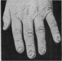
Fig. 240.—Atrophy of the nails.
1Longstreth, “On the Changes in the Nails in Fever, etc.,” Trans. Coll. Physicians
of Philadelphia, 1877, p. 113.
2 Wilks, Trans. Pathol. Soc’y, London, 1870, p. 409.
3 Hartzell, discussion on Disease of the Nails, Trans. Amer. Derm. Assoc. for 1901.
4 Zeisler, “Trophic Dermatoses Following Fractures,” Jour. Cutan. Dis., 1898, p.
305.
5 Crocker, Diseases of the Skin.
ATROPHIA UNGUIUM 959
times the toe-nails also, although this latter is not so frequent. It may
begin in any portion—ordinarily, however, at the basal portion. The
nails break and crack readily. Poor health, chronic digestive disturb
ances, diseases of the nervous system, possibly traumatisms, and invasion
by the vegetable fungi of favus and ringworm are factors in different
cases.
Shedding of the nails occurs sometimes after fevers and nervous
diseases. It has been occasionally observed conjointly with alopecia
areata and general defluvium capillorum. It is also seen in diabetes,
scarlatiniform erythema, and dermatitis exfoliativa, but in other in
stances without apparent cause. In some cases it is hereditary and even
congenital. Montgomery1 reported a case of a man in whom there had
been a constant shedding of the finger-nails since birth, with a history
of a similar affection in some of his ante
cedents. Apparently in certain nervous
disorders, diabetes, etc, the great toe-nail
most frequently suffers in this respect.
Leukopathia unguium (achromia un-
guium; leukonychia; flores unguium;
white spots; white nails; gift spots,
etc), in its mildest type, is not infre
quent, the chalky whiteness being either,
and most usually, in the form of spots
or in the form of transverse bands, the
nails otherwise being quite normal.
They appear near the lunula, and gradu
ally move forward with the growth of
the nail. Unna,2 Giovannini,3 Long-
streth,4 Morison,5 Stout,6 Heidingsfeld,7
and others report marked instances of
the band type; Longstreth observed
white transverse bands on his own finger-
nails after an attack of relapsing fever;
the several bands marking the time of the relapses. Giovannini’s case
followed typhoid fever, and Unna’s case was apparently congenital, and
associated with partial ringed hair. Morison’s patient, a young woman,
was in good health, the bands appearing without apparent cause; they
had practically disappeared for a time one summer. In Stout’s case, a
mulatto, in addition to involvement of the finger-nails, a number of the
toe-nails exhibited a similar, but slightly less marked, condition; and so
1D. W. Montgomery, Jour. Cutan. Dis., 1897, p. 252 (with some literature
references).
2 Unna, International Atlas, plate xix, 1891.
3 Giovannini, ibid.
4 Longstreth, loc. cit.
5 Morrison, Archiv, 1888, p. 3 (with colored plate).
6 Stout, Medical News, Feb. 24, 1894 (with illustrations and some literature refer
ences) .
7 Heidingsfeld (7 cases), Jour. Cutan. Dis., 1900, p. 490 (with illustrations and
bibliography); Sibley, Brit. Jour. Derm., 1911, p. 281, records a case, and also a case
of yellow-ochre color in a syphilitic subject while under antisyphilitic treatment (with
a review of the literature with references).

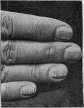
Fig. 241.—Leukopathia ungui-
um (the transverse band type);
patient manicured herself about
every seven to ten days.
960 DISEASES OF THE APPENDAGES
far as could be ascertained was probably congenital. Lawrence,1 quoted
by Stout, observed an example of finger-nail bands in a healthy man aged
forty-five, apparently congenital, and whose child, aged five, likewise
presented a similar appearance, though not so distinctly marked.
Etiology and Pathology.—The causes of these various atrophic
and other described conditions have in part been incidentally referred
to. So far as observation goes, the same etiologic factors responsible
for the production of hypertrophy may also lead to atrophic changes.
The various inflammatory, and especially scaly, skin diseases, more
particularly when involving the hand and fingers, are often etiologic;
constitutional diseases, nervous disorders, glossy skin (Weir Mitchell),
traumatism, vegetable parasites, etc, are therefore variously found in
the different cases; as already stated, heredity is a demonstrable factor
in some of the cases. In many, however, it must be confessed, it is
difficult to find an adequate explanation for the often persistent and
rebellious atrophic manifestation. The immediate factor, exclusive
of the parasites, is doubtless trophic in character, but that is about
as far as one can get in many instances, and that is more of a cloak for
lack of knowledge than a satisfying explanation. Nutritive disturbance
of the matrix naturally is followed by imperfect nail-formation, and
this part shares in all general enfeebling diseases, especially if profound
and long continued.
The white spots and bands are in most cases either due to general
disease, such as fevers, nervous disorders (Bielschowsky) ,2 or to local
traumatisms. In many instances, however, as in that of complete in
volvement in Joseph’s3 patient, there is no assignable cause. The striated
form is rare, although Heidingsfeld’s observation of 7 well-marked cases
in a comparatively short time indicates that it is not so rare as it ap
parently seems from the scant literature of the subject. Six of his
patients were young women who assiduously manicured their nails, and
his studies would indicate that the cuticle knife is a possible cause of these
white formations. He gives an illustration of 1 case, showing the nails
one-half (new-growing part) normal after disuse of the cuticle knife for
forty days. As yet there is some difference of opinion as to the origin or
production of the white appearance. Most of the various writers named
and other observers consider it due to infiltration of air, filling the minute
interstitial spaces between the loosened-up epithelial strata. This is the
accepted view. Heidingsfeld, who made careful microscopic examina
tions of the affected nails, was not able to corroborate this, and he con
sidered that his researches justified the following: Leukoplakia unguium
is the result of some pathologic change of structure of a plane of nail-
cells, approximating a failure of the affected cells to undergo normal
physiologic keratinization; the causes may be trauma, malnutrition,
febrile diseases, neuroses, or any agency which disturbs the growth,
development, or keratinization of matrix cells in their change to nail
1 Lawrence, Australian Med. Jour., Oct. 15, 1893.
2 Bielschowsky (following multiple neuritis), Neurologisches Centralblatt, 1890, p.
741.
3 M. Joseph, “Leukonychia totalis,” Dermatolog. Zeitschr., 1894, p. 657.
ONYCHOMYCOSIS
96l
structure; an infiltration of air is not present, and there is no rational
physiologic basis for such a theory.
Treatment.—The management, in a general way, of nail atrophy
is essentially the same as described in treatment of hypertrophy of these
structures. A recognition of the causative factor is important for suc
cess, but this often seems impossible. In the absence of underlying
factors which will give indication of constitutional treatment when
necessary an empirical plan is the only resort. Arsenic, and also cod-
liver oil, often have a favorable influence if persevered in. Small doses—
2 or 3 grains (o. 135-2.) three times daily—of sulphur in some instances
appear to have an alterative effect. The local management consists
in the protection of the parts from traumatism and from contact with
disturbing materials, such as water, other liquids, and irritating sub
stances. When necessary, disinfection with boric acid solution and appli
cations of the milder salves, such as prescribed in eczema, and those named
in the treatment of nail hypertrophy are useful. In cases of nail-splitting,
enveloping the part with salve nightly, and wearing over the finger-end
a piece of a glove-finger, and keeping the free end of the nail closely cut
or filed, will, if persisted in, often get rid of the trouble.
In white nails the treatment is purely upon general indications,
with a trial of arsenic in chronic cases, and, if necessary, the conceal
ment of the blemishes by some indifferent stain; for this latter purpose
occasionally touching the spots with a 5 or 10 per cent, resorcin lotion
will bring about slight discoloration and render the blemish less conspicu
ous. The treatment of atrophic nails due to the ringworm and favus
fungi will be elsewhere considered (see Onychomycosis).
But first, if you want to come back to this web site again, just add it to your bookmarks or favorites now! Then you'll find it easy!
Also, please consider sharing our helpful website with your online friends.
BELOW ARE OUR OTHER HEALTH WEB SITES: |
Copyright © 2000-present Donald Urquhart. All Rights Reserved. All universal rights reserved. Designated trademarks and brands are the property of their respective owners. Use of this Web site constitutes acceptance of our legal disclaimer. | Contact Us | Privacy Policy | About Us |