| MEDICAL INTRO |
| BOOKS ON OLD MEDICAL TREATMENTS AND REMEDIES |
THE PRACTICAL |
ALCOHOL AND THE HUMAN BODY In fact alcohol was known to be a poison, and considered quite dangerous. Something modern medicine now agrees with. This was known circa 1907. A very impressive scientific book on the subject. |
DISEASES OF THE SKIN is a massive book on skin diseases from 1914. Don't be feint hearted though, it's loaded with photos that I found disturbing. |
EPITHELIOMA
Synonyms.—Skin cancer; Epithelial cancer; Carcinoma epitheliale; Cancroid;
Noli me tangere; Fr., Epithéliome; Cancroide; Ger., Hautkrebs; Epithelialkrebs.
Definition.—The term epithelioma, in its strict dermatologic
significance, may be defined as an epithelial new growth with destructive
tendency, having its origin in the epithelium of the epidermis or of the
glands of the skin.
Symptoms.—Epithelioma presents, when at all developed, va
rious clinical phases or characters, usually with, in different cases, a
predominance of one of the several features over the others, so that it
would be possible to make a number of varieties. For practical and
descriptive purposes, however, the three divisions commonly made are
1Milligan, Brit. Jour. Derm., 1911, p. 411 (case demonstration—patient, woman
aged thirty-one).
CARCINOMA CUTIS 871
sufficiently comprehensive. These several forms of epithelioma are desig
nated the superficial, or discoid, the deep-seated, and the papillomatous.
Superficial Variety.—The superficial, or discoid, form, sometimes
also named the flat variety, is, of the several varieties, that most fre
quently encountered. It may begin in one of several ways: as a small,
firm, reddish, yellowish, or pearly papule or tubercle, or as an aggrega
tion of two or three such lesions; as a warty excrescence, primarily of a
benign character, such as a simple wart or small, elevated, fleshy mole;
and as a small pea- to bean-sized, smooth, or roughened keratosis or
greasy, scaly, seborrheic patch (see Keratosis senilis and Atrophia senilis).
In the event of it beginning as one of the several small, solid lesions named,
the first evidence of the degenerative change is usually a slight fissure or
surface abrasion, which persists and slowly enlarges, and often becomes
thinly crusted. After some months the slight pearly, warty, or mole
formation has in great measure slowly yielded to the degenerative action,
and in great part or wholly disappeared, giving place to a surface ulcera-
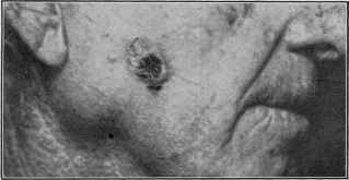
Fig. 212.—Epithelioma, superficial type, in an old man aged sixty-five, showing the
roll-like border.
tion a trifle larger than the lesion which it has gradually displaced. In
some cases, however, a long time elapses—some months or one or two
years—before the first lesion has wholly gone, and it is not uncommon
for it to be gradually destroyed in the upper part, and the basal portion
remain for a shorter or longer period, with an ulcerated surface covered
with a thin or slightly thickish incrustation, which is knocked off or falls
off from time to time, but which soon reforms. After the lesion has,
however, sunken to and usually a trifle below the skin level and a shallow
ulcer is in its place, it is commonly observed to have a slightly elevated
and often a pearly, roll-like border, which is gradually pushed further
as the ulcer becomes larger. In rare instances, as in those observed by
Danlos, Brocq, Fordyce, Hartzell, and myself,1 the superficial epithe-
1 Danlos, Annales, 1899, p. 656 (case demonstration—patient, woman aged forty-
three, with lesion on the neck); Brocq, ibid, (discussion—2 cases, both women, aged
fifty-five and sixty, with lesion about the nose); Stelwagon, Trans. Amer. Derm. Assoc.
for 1899, p. 166 (case demonstration—patient, male, aged forty, with flat epithelioma
of the temporal region, with almost an inch-wide superficial, morphea-like border);
Fordyce, Jour. Amer. Med. Assoc., Oct. 24, 1908, p. 1398; Hartzell, “Morphea-like
Epithelioma,” Jour. Amer. Med. Assoc, July 24, 1909, p. 262 (3 cases with brief
review and references).
872
NEW GROWTHS
lioma spreads by an invading flattened, morphea-like, slightly raised
band of 1/4 to 1/2 inch or so in width (morphea-like epithelioma); the
morphea-like formation, but slightly raised, seems to be the first stage,
this later showing trifling crusting with underlying degenerative changes
in the central portion; the ulcerative action slowly spreads, and the
morphea-like border at the same time may also extend.
The ulcer, which has a slight serous or serosanguinolent, viscid,
or varnish-like discharge, keeps more or less continuously crusted,
and the crust, when it is not frequently rubbed or knocked off, may
become quite thick. The discharge is sometimes mixed with pus, but
it is only, as a rule, in the more advanced stages, when the ulcer becomes
large and tends to extend more deeply, that the purulent character of
the discharge is at all noticeable, and never so pronounced or distinctly
purulent as in syphilitic ulcers. The superficial variety of epithelioma
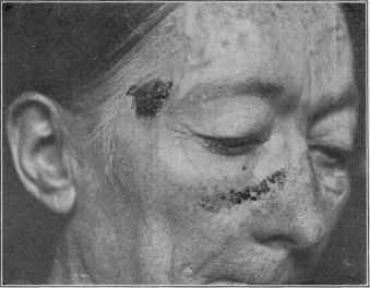
Fig. 213.—Epithelioma of superficial type, crusted, in a woman aged fifty-eight.
rarely, certainly not, as a rule, until after some years’ duration, extends
to much depth. In course of time it does, it is true, gradually eat in,
so that when on the side of the nose there is risk of final penetration.
In occasional instances of these sluggish superficial types, and especially
that form with the pearly border, as the disease spreads there is displayed
in the older parts a tendency to cicatricial healing.
In other cases the disease pursues a course which has given it the
name of rodent ulcer (Jacob’s ulcer; Cancroid ulcer; Ulcus exedens:
Ulcus rodens; Noli me tangere; Ulcére cancreux; Der flache Hautkrebs)1,
formerly these cases were also designated lupus exedens. The special
characteristic of this type is its lateral, steadily progressive spread, with
but little, and sometimes no, elevated or infiltrated border; in other
words, the ulcerating feature is conspicuous, whereas the new growth
element is almost nil. Not infrequently, especially in the earlier stages,
CARCINOMA CUTIS
873
there is a slight, pearly, roll-like border. Some observers are inclined
to consider this a distinct malady from epithelioma, although admitting
it to be an allied disease, but the origin, behavior, and pathologic charac
ters are in all essential particulars similar to those of other superficial
cases. It is true, as Paget states, that it frequently begins as a brownish
nodule, different from the pearly tubercle or warty growths which mark
the other superficial type, but rodent ulcer, or a lesion clinically indis
tinguishable, may also begin, as I have often observed, in the same man
ner as in the cases of the superficial form already referred to. The rodent
ulcer variety is commonly a disease of the upper half of the face, being
especially frequent about the eyelids and sides of the nose.
The course of superficial epithelioma, as has already been intimated,
is usually pre-eminently slow, and the disease of a relatively benign char-
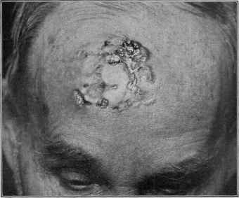
Fig. 214.—Epithelioma, rodent ulcer type, in a man aged sixty, of fifteen years’ dura
tion; recurrence several times after curetting and cauterization.
acter, many years often elapsing before serious progress has been made.
The rodent ulcer form is often eventually extremely destructive, extend
ing deeply as well as laterally. The lymph-glands are rarely involved
in these superficial cases, but there is a possibility in all instances of final
glandular involvement1 and a change of type into a deep-seated or papil
lary variety of the disease.
Deep-seated Variety.—The deep-seated or nodular variety of epi-
thelioma may start from the superficial type, or it begins as a tubercle
or nodule in the skin or subcutaneous tissue. It gradually increases
in dimensions, projecting both downward and above the level of the skin,
with the overlying integument pinkish or reddish, and frequently with
1 See interesting paper by D. W. Montgomery, “Report of a Case of Epithelioma
of the Skin with Unusual Course of Infection of Lymph-nodes,” Annals of Surgery,
1898, vol. xxvii, p. 193.
874 NEW GROWTHS
dilated capillaries coursing over it. There may be some lateral extension
into the surrounding tissue. In the course of several months or longer
the nodule, which has frequently reached the size of a cherry or larger,
breaks down centrally, an ulcer is formed, with usually prominent and
infiltrated reddish and inflammatory-looking borders. The surface
of the ulcer is reddish and granu
lar and secretes a viscid, and often
ichorous discharge, and crusts
over from time to time. When
arising from the superficial variety
of epithelioma, this latter is
usually noted to become more
angry-looking, especially about
the edges, the latter becoming
more prominent, showing increas
ing infiltration, and the base of
the growth likewise tending to be
come thicker and hard, and there
may, in some instances, be considerable elevation. The progress of the
deep-seated type is usually steadily progressive, and, as a rule, at a rela
tively rapid pace. The infiltration, which is the pronounced feature of
this form, spreads gradually, sometimes rapidly; the ulcer enlarges both
peripherally and in depth, and presents hard, everted, often more or less

Fig. 215.—Small beginning epithelioma.
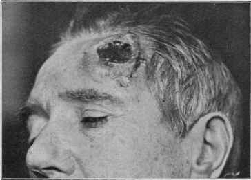
Fig. 116.—Epithelioma, deep-seated, “crateriform” variety.
undermined edges, sometimes showing here and there small waxy nodules;
the base is noted to be irregular, not infrequently slightly or moderately
papillomatous or vegetating, and with often considerable viscid, varnish-
like, sanguinolent, partially or markedly purulent discharge. The base
exhibits, as a rule, a disposition to slight bleeding upon the slightest
CARCINOMA CUTIS 875
provocation, such as a trifling knock or insignificant roughness in its
washing or dressing. In some cases the ulcerative tendency is displayed
chiefly in the central portion, the surrounding infiltration being somewhat
hard and elevated, and the invasion and progress rapid, constituting the
“crateriform ulcer” of Hutchinson.1 This rare variety, which usually
is seated on the upper part of the face, may also start as a superficial
rodent ulcer type. Muscle, cartilage, and bone often finally become
invaded. The neighboring lymphatic glands are sooner or later im
plicated; pains of a burning or neuralgic type are experienced, and with
or without recognizable, metastatic tumors in the internal organs, death
eventually ensues.
Papillary Variety.—The papillary or papillomatous variety of epithe-
lioma usually arises from the superficial or deep-seated type; or it may
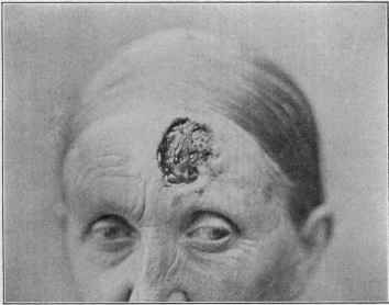
Fig. 217.—Epithelioma of deep-eating rodent type, in a woman aged sixty (courtesy of
Dr. A. Van Harlingen).
begin primarily as a papillary or warty growth. Beginning in the latter
manner, it may for some months or longer maintain a pseudoverrucous
appearance, projecting higher, however, than ordinary warts, involving
more surface,—1/2 to 1 inch or so in diameter,—with a slightly or
moderately, mildly inflammatory, infiltrated base, which may extend a
line or two beyond the edge of the papillary formations. When the area
of disease is small, the vegetations are usually noted to be somewhat
higher centrally, although in larger areas also the papillary projections
are generally much less prominent toward the extreme peripheral portion.
When a papillary epithelioma is fully developed, it matters not in what
manner it may have originated, it presents an ulcerated, fissured, and
papillomatous surface, usually having a viscid or thick secretion, with a
1 Hutchinson, “The Crateriform Ulcer of the Face, a Form of Acute Epithelial
Cancer,” Trans. London Patholog. Soc’y, 1889, vol. xl, p. 275 (with colored plates).
876 NEW GROWTHS
variable proportion of purulent admixture. The surface may bear re
semblance to a digitate wart, to a cauliflower excrescence, or it may be
distinctly condylomatous in appearance. Exceptionally there is a
tendency toward slight pedunculation. Sometimes the secretion is
scanty, the ulcerative action more in the nature of deep fissures extending
down between the papillary projections, and the surface of the growth
may present a somewhat hard or horny, thin or moderately thick incrus
tation. In some cases the surface is irregularly ulcerated and papillo-
matous, the granulations usually presenting an exuberant and fleshy
character, which bleed quite readily. It is slowly, sometimes rapidly,
progressive, and sooner or later develops a malignant tendency, showing
deep-seated infiltration, involvement of the neighboring lymph-glands,
and death, as in the malignant, deep-seated variety, gradually results.
Epithelioma of the lip, usually on the lower lip toward one side,
comes more commonly under the care of the surgeon, although in its
earliest stage it is not infrequent in dermatologic practice. It begins in
one of the several ways described in connection with its appearance on the
cutaneous surface. Its most common commencement is as a slight scurfi-
ness, abrasion, crack, or small papule or warty-looking lesion, and in many
cases advice is not sought until a superficial ulcer, with variable infiltra
tion, is noticed, which presents in several months or longer. The future
course is about the same as with the more rapid skin cancers, there being
usually considerable infiltration and swelling, with comparatively early
involvement of the lymphatic glands. The surface is commonly granular-
looking, in places often crusted. Cancer of the tongue, which may also,
in its beginning stage, first come under the inspection of the dermatolo
gist, begins as a small abrasion or fissure, often started by irritation pro
duced by a tooth, or developing upon a leukoplakia. A superficial ulcer
soon results, later infiltration beneath and surrounding, and more or
less rapid course and destruction. Epithelioma of the genital organs is
also met with, the glans penis, prepuce, and clitoris being not unusual sites.
On these parts the papillomatous variety is of common occurrence,
starting from an abrasion or a warty growth.1
The favorite sites of election in skin cancers are, first of all, the various
parts of the face, especially the nose, eyelids, and lips. The forehead
and, more frequently, toward or at the temporal region, the ear, the back
of the hand, and the genitalia are also localities frequently invaded.
Any other part may, however, be, but, as a rule, only rarely, the seat of
such growth. On the dorsal surface of the hand the lesion begins usually
either as a warty excrescence, developing into the papillomatous type,
or as a keratosis or degenerative seborrheic patch, which scales or crusts
off from time to time, as already described, and gradually breaks down
and into the ordinary epitheliomatous ulcer. As a rule, the growth is
single, although in exceptional cases two or three lesions may be present;
and in some instances of numerous, scattered, senile, degenerative, sebor-
rheic patches about the face, several or more may gradually undergo
epithelial change,—épithéliomatose sébacée of the French,—and mul-
1 For detailed description and management of lip, tongue, and genital cases the
reader is referred to works on surgery.
CARCINOMA CUTIS
877
tiple epithelioma result. Mention should also be made here of the con
dition occasionally met with, which will be found described elsewhere
under the title “multiple benign cystic epithelioma” (q. v.). In this,
numerous lesions, discrete or bunched, usually about the face or upper
part of the trunk, are present, of a character closely similar, if not identi
cal with, the pearly growths and border, both clinically and pathologic
ally, seen in some cases of ordinary epithelioma; in view of the few in
stances in which in this mild malady degenerative changes have occurred
it is highly probable that the future of these cases may show that the
term “benign” is scarcely acceptable.
In the milder phases of epithelioma no subjective symptoms are
complained of, but in the deep-seated and papillomatous varieties
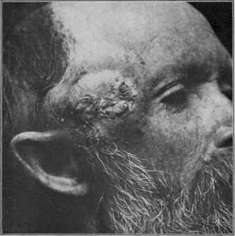
Fig. 218.—Epithelioma of deep-seated type in a man aged fifty-seven.
burning sensations or pain of a lancinating character frequently develop
late in the disease. Unless in close proximity to lymphatic glands,
these, in average cases, rarely show involvement in the earliest months,
and sometimes never during the whole course of the malady; but if
closely situated, more especially with the deep-seated and papillomatous
varieties, the lymphatics are sooner or later implicated. In the average
run of cases of the type naturally gravitating to the dermatologic special
ist, however, glandular implication, judged by my own observation,
which covers a large aggregate number, is relatively rare; nor have I
seen, with the exception of a few instances, a recurrence presenting itself
in the glands. It is only fair to state, however, that most of my cases,
878 NEW GROWTHS
fortunately, have happened to be of the mild, superficial variety, with
only in a moderate proportion of instances a disposition to a rapidly
malignant tendency.
Etiology—The cause of cancer is still an undetermined question.
There are several factors, however, which may be considered as adjuvant
or contributing that are well known. The majority, by far, of epithelio-
mata of the skin or adjoining mucous surfaces are observed in males.
The malady is essentially one of advancing years, somewhat rare before
the age of forty, and more commonly seen after fifty or sixty. Excep
tional instances, it is true, are now and then noted, as in the recent cases
recorded by Hartzell,1 Allen,2 Sequeira,3 and others, in which the growth
presents in earlier years. Local irritation is likewise a recognized ex-
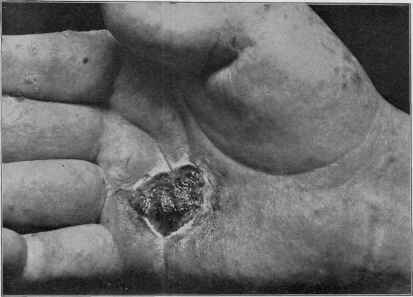
Fig. 219.—Epithelioma developing from a keratosis, in a case of psoriasis; the
keratoses (some of which can be seen in the illustration) appearing after long-continued
administration of arsenic.
citing agent. This is shown in the cases in which, from the accidental
knock or other injury, often trifling, an ordinary wart, fleshy mole, or
pigmented nævus of long duration begins to show epithelial development.
The disease has also often been noted to start at the site of a scratch,
cut, or other accidental traumatism; and the factor of pressure or irrita
tion produced by the pipe-stem or cigar in the production of epithelioma
1 Hartzell, “Epithelioma (Rodent Ulcer) in a Boy of Fourteen,” New York Med.
Jour., Mar. 5, 1898 (refers to several recorded cases, with literature references).
2 Allen, Jour. Cutan. Dis., 1899, p. 571 (case demonstration, in man aged twenty-
four, on the lip); ibid., 1900, p. 122 (case demonstration—patient male, aged twenty-
eight—on lip).
3 Sequeira, Brit. Jour. Derm., 1912, p. 391, reports a case of “Rodent Ulcer of the
Back in a Boy of Twelve,” and refers to his previous cases—one on the ala nasi, begin
ning when the patient was twelve, and another on the lower lid of a girl aged fifteen.
CARCINOMA CUTIS 879
of the lip in many instances is well known. A neglected or irritated
senile seborrheic spot or keratosis often is, as already referred to, the
starting-point of the disease. Hyde,1 Dubreuilh,2 and others have called
attention to the possible factor of continued exposure to the sun’s rays;
and it is well known that workers in petroleum and tar products develop
keratoses and papillomata with malignant tendency. Epithelioma has
also developed at the site of patch or ulceration of lupus vulgaris and
syphilis, and exceptionally upon a lupus erythematosus. Hutchinson,
White, Hartzell, and others have recorded cases developing, in psoriasis
patients, from the keratosis following the prolonged administration of
arsenic,3 this drug, therefore, having apparently a direct—certainly
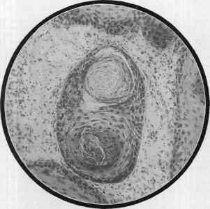
Fig. 220.—From a squamous-celled epithelioma, showing the so-called “pearls,” “cell-
nests,” or “globes” (courtesy of Dr. J. A. Fordyce).
indirect—influence (arsenical cancer, arsenical epithelioma). From
time to time various claims as to the discovery of parasitic organisms
have been made, but a judicial review of the evidence shows that this
field of investigation has not, up to the present, borne convincing results.
So far we have not got beyond the recognition of a local irritation—espe-
1 Hyde, Amer. Jour. Med. Sci., January, 1906.
2 Dubreuilh, Annales, 1907, p. 387 (with valuable statistical tabulations of
epithelioma cases from Ferrer’s “These (Bordeaux, 1906-07), Etiologie clinique de
l'epithelioma cutané” (432 cases).
3 Hartzell, “Epithelioma as a Sequel of Psoriasis and the Probability of its Arsen
ical Origin,” Amer. Jour. Med. Sci., Sept., 1899 (report of a case and review of
recorded cases, with references); Dubreuilh, “Keratose arsenicale et Cancer arsenical,”
Annales, Feb., 1910, (adds few cases, and reviews the subjects of arsenical keratosis and
arsenical cancer with tabulations of reported cases, with references); Wile, “Arsenical
Cancer, with a Report of a Case,” Jour. Cutan. Dis., 1912, p. 192 (on lingers; good re
view, with references; 19 cases collected).
88o
NEW GROWTHS
daily of a keratotic lesion and frequently of other benign skin lesions—
being a factor of importance; and that this irritant may be of various
kinds and sources1
Pathology.—Histopathologic studies of the epitheliomatous proc
ess which especially interests the dermatologist, show that it consists,
briefly described, in the proliferation of epithelial cells—pavement
epithelium—from the epidermis or from the epithelium of the hair-
follicles or glandular structures,2 or from the mucous membrane; the cell-
growth takes place downward, in the form of finger-like prolongations or
columns, or it may spread out laterally and deeply so as to form rounded
masses, the centers of which usually undergo horny transformation,
resulting in the formation of onion-like bodies, the so-called “pearls,”
“cell-nests,” or “globes.” The rapid cell-growth requires increased
1 In a paper in which the possible etiologic factors are gone over, and carefully
considered, Hartzell (“Etiology and Pathology of Malignant Diseases of the Skin
Affecting Epithelial Tissue,” Jour. Cutan. Dis., 1900, p. 435), concludes as follows:
“We may regard it as fairly well demonstrated that carcinoma results from a pro
found and more or less permanent alteration of the mechanism of cell-division. This
alteration may, in my opinion, result from long continued irritation of a mechanical or
chemical kind, including under this latter the effects of toxins resulting from micro-
organisms. Accordingly it seems likely that the immediate causes of cancer are
multiple.”
Ten years later, in a similar study and review, Fordyce (“The Pathology of
Malignant Epithelial Growths of the Skin,” Jour. Amer. Med. Assoc, Nov. 5, 1910,
p. 1624) reaches practically the same conclusion: “A study of skin cancers suggests
to the observer, if it does not demonstrate absolutely, that no one agent is con
cerned in the malignant proliferation of epithelial tumors and that cutaneous car-
cinomata have a multiple etiology. The development of epitheliomata following
exposure to sunlight, x-rays, or other radiant energy is a strong argument against
the parasitic nature of the disease. Likewise, the occurrence of epitheliomata in
xeroderma pigmentosum and allied conditions of the skin which come on in old
age or middle life is an additional argument against this theory. These conditions
are preceded by changes identical with those met with in xeroderma pigmentosum,
such as a dry atrophic skin, telangiectases, warty growths and, finally, malignant
transformation. Furthermore, the action of chemical substances on epithelium, for
which they have a special predilection, such as arsenic, tar, scarlet R., tobacco, etc.,
demonstrate that a variety of agents have the power to stimulate epithelial mitoses
which may pass into malignancy. Cancers which develop on scar tissue or antecedent
conditions of the skin like lupus, syphilis, etc, suggest that we are dealing with mis
placed cells in some cases and in others with degenerative processes which lead to the
abolition of the functional activity of the cells, which is followed, as a consequence,
by vegetative activity, according to the theory of Oertel, Adami, and others. In
primary multiple epitheliomata we have several foci in which an infectious agent or
some internal sensitizing agent may have acted on the cells and rendered them sus
ceptible to a local factor.”
See also paper by Schamberg, “Cancer in Tar Workers,” Jour. Cutan. Dis., 1910,
p. 644 (4 personal cases; review of the literature bearing upon cancer in workers in tar,
paraffin, soot (chimney-sweep’s cancer), with bibliography); by Loeb, “Ætiology of Can
cer of the Skin,” Jour. Amer. Med. Assoc, 1910, lv, p. 1607 (reviews the various theories
and concludes that irritation is of the greatest etiologic significance); Bowen, “Precan-
cerous Dermatoses,” Jour. Cutan. Dis., 1912, p. 241 (report of 2 cases; reviews the
subject of the various precancerous dermatoses with literature references); Sachs,
Wien. klin. Wochenschr., Nov. 9, 1912 (remarkable production of warts and warty
eczema on the hands of those working in anilin dyes; experimental investigations with
animals (rabbits) confirmed the property of these dyes to induce granulation and epi-
thelioma-like excrescences, which may undergo degeneration).
2 As to its origin in the sweat-glands see Fordyce’s paper, “Adenocarcinoma of the
Skin, Originating in the Coil-gland,” Jour. Cutan. Dis., 1895, p. 41 (report of a case
with histologic cuts and review and references), and for an admirable general pres
entation with case illustrations and histologic cuts; Fordyce, “Clinical and Patho
logical Observations on Some Early Forms of Epithelioma of the Skin,” New York
Med. Jour., June 9 and 23, 1900.
CARCINOMA CUTIS
881
nutriment, and hence the blood-vessels become enlarged; moreover, the
pressure of the cell-masses and their invasion of other tissues give rise
to irritation and inflammation and consequent increased blood-supply,
with corresponding serous and round-cell infiltration.
Epitheliomata of the skin histologically present two types, the lobu-
lated and the tubular. The former, which is the more frequent, shows,
as the name signifies, a massing of the epithelial new growth in the form
of lobules, and each lobule is noted to be composed of concentric strata
of cells, which correspond in their changes to the several strata of the
epidermis—from those of the rete to those of the corneous layer. The
Fig. 221.—Epithelioma—section from the margin of a deeply eating rodent ulcer
variety of the eye region; eye already destroyed (courtesy of Dr. A. R. Robinson).
innermost cells of the lobule show imperfect cornification, while the outer
most cells correspond to those of the basic cylindric cells of the rete,
those of between layers showing the various changes from the latter to
the former. Owing to the pressure upon the central mass of cells, the
onion-like bodies, or cell-nests, already referred to, are produced. These
are usually found, at least most abundantly and of typical formation,
in the deeper parts. In some instances this central portion undergoes
colloid degeneration. Offshoots from the down-growing lobules are
frequently noticed, and these present the same characters and undergo
the same changes as in the parent lobules. When the epithelial masses
56
882 NEW GROWTHS
are of rapid growth, the cells, from mutual pressure, may, in places, be
spindle shaped. In the tubular or cylindric type the epithelial growth
is in the form of cylindric processes, which freely anastomose with one
another. They run usually more or less perpendicularly to the sur
face, but in some instances may be parallel to the epidermis and occa
sionally presenting a pseudoglandular appearance. The cells are com
posed of smaller cells than those of the other variety, and correspond
more closely to those of the deeper layer of the rete. They show prac
tically no tendency to horny transformation and the formation of the
cell-nests, which occur in the lobulated types of growth.
The conditions in the rodent ulcer type are essentially those of
tubular epithelioma, many observers believing that the epithelial pro
liferation takes its origin in the epithelium of the rete (Collins Warren,
Robinson, Unna),1 of the hair-follicle (Tilbury Fox, Colcott Fox, Sang-
ster, Hume),2 sweat-glands (Thin, Walker),3 sebaceous glands (Thiersch,
Butlin). Fordyce4 views it as a small-celled epithelioma originating
from the deep layer of the epidermis or the hair-follicles. Paul, Boyce,
Darier, and others regard it simply as a slow-growing epithelioma which
may start from the epithelium of any of the skin structures, and this view,
judged by the various findings referred to, is probably the correct one.
Bodies thought to be organisms, supposed to be coccidia, have been
found in cancer-cells, chiefly by Albarran and Malassez; they occur in
very small number, and principally in the very center of the cell-nests,
but the investigations and studies of Borrel5 and Hutchinson,6 Jr., have
led them to the conclusion that they are not of parasitic nature. Welch,
Noeggerath, Török, and others (cited by Fordyce), have also failed to
substantiate the claims. Gaylord’s interesting observations as to the
presence of organisms are too incomplete to warrant a definite conclu
sion. Most of the findings so far made by investigators are thought to
represent various forms of cell degeneration.
Diagnosis.—The main diagnostic points in epithelioma to re
member are: the age of the patient; the usually single character of the
growth; its beginning in a wart, mole, nodule, or scurfy spot; the char
acter of the border—pearly, with roll-like elevation or a hard, elevated
infiltration; the scant, and, in the later stages, viscid discharge, frequently
streaked with blood; its usually slow progress; the frequent situation
about the nose, eyelids, or other parts of the face; its finally involving,
in many instances, the neighboring lymphatic glands, more especially
when the lesion is seated near these structures. Microscopic examina
tion of a section of the tissue is generally conclusive in doubtful cases.
1 Collins Warren, The Anatomy and Development of Rodent Ulcer, Boston, 1872;
Unna, Histopathology.
2 Tilbury Fox and Colcott Fox, London Patholog. Soc’y Trans., 1879, vol. xxx, P.
360 (with histologic cuts); Sangster, Brit. Jour. Derm., 1882, p. 777.
3 Thin, loc. cit.; Walker, Brit. Jour. Derm., 1893, p. 286.
4 Fordyce, loc. cit., and Morrow’s System, vol. iii (Dermatology), p. 655, to whose
article I am indebted.
5 Borrel, “Sur la signification des figures décrites comme coccidies dans les épithé-
liomes,,, Arch, de méd. exper., 1890, p. 786 (with colored plate presenting 5 histologic
cuts).
6Hutchinson, Jr., “On Psorosperms and Skin Diseases,” Brit. Jour. Derm., 1891,
p. 277.
CARCINOMA CUTIS 883
The lesions from which it is to be differentiated are syphilitic ulcera-
tion, warts, and lupus vulgaris. The differentiation from the last is
given under that disease. The tubercular ulcerating syphiloderm,
like epithelioma, is frequently seated upon the face, but it differs in sev
eral particulars from the latter. It is usually multiple, consisting of
several superficial ulcerations and not, as a rule, rounded in shape, but
segmental or irregularly circinate. Tubercles which have not as yet
undergone destructive change, or without such tendency, are also com
monly seen in association with the syphilitic ulcerations, and with the
same disposition to the segmental or serpiginous configuration. More
over, syphilitic ulcers have, as a rule, quite a free discharge, and generally
of distinctly purulent character.
Benign, warty-looking formations and fleshy moles are to be differen
tiated from those of beginning malignant growths by attention to their
history and course; in fact, long-continued observation may be necessary
before a positive opinion is warrantable. The appearance of any tend
ency to crusting, to break down, or ulcerate, is significant of epithelio-
matous degeneration. Such a benign-looking lesion, showing, therefore,
one or two fissures at the summit or edge of the base, or exhibiting slight
abrasions, which are persistent, is to be viewed as a beginning epithe-
lioma.
On the lip a persistent, localized thickening or abrasion of several
months’ or more duration, especially when on the lower lip, and occurring
in an individual over forty, means almost always an epithelioma. The
possibility of this region being the seat of the initial lesion of syphilis is
not, however, to be forgotten, and in the earliest stage a differentiation
is sometimes impossible, but the more rapid development of the latter,
with usually but little tendency to active or large ulceration, and with
soon the appearance of secondary symptoms of the disease, will serve
for final differentiation. About the genitalia epitheliomata practically
present the same features as elsewhere, frequently beginning as a warty-
looking lesion, and slowly and gradually developing into a spreading
infiltrated and destructive epitheliomatous ulcer. It is here to be dis
tinguished from a chancre and from the tuberculogummatous infiltration
and ulceration of later syphilis.
Prognosis.—In an opinion as to prognosis in epithelioma several
factors are to be considered—the variety, extent, duration, and rapidity
of the process. In all instances the earlier treatment is instituted, the
less chance is there of recurrence. In many cases of the more superficial
forms, the disease, even if neglected, is slow in its progress, often lasting
for ten, fifteen, or more years before seriously threatening the patient’s
life. Thin1 records an extreme instance of a cancerous ulcer of the rodent
type in a woman aged sixty-eight, involving shoulder and upper part
of the back, which had begun forty-three years previously as a pimple
or wart. In the earliest stages of the cases ordinarily met with in derma-
tologic practice, when the disease is limited, on the face, and of a super
ficial type, treatment is almost invariably successful and permanently
1 Thin, London Patholog. Soc’y Trans., 1879, vol. xxix, p. 237 (was histologically
of cylindric type and had apparently started in the sweat-glands).
884 NEW GROWTHS
so. I have had a large number of such cases under my care, and if not
far advanced or if not of the rapid, deep-seated, or papillomatous type,
with no glandular involvement, the result has been uniformly good.
Even when such cases are moderately advanced, the outlook is usually
favorable. The same may be said of the deep-seated and papillomatous
varieties if not of too long duration, but in these glandular involvement
is, after a time, not uncommon, and quite the rule later. Cases in which
conspicuous destruction has already ensued, and in which there is con
siderable surrounding infiltration and of long duration, the prognosis as
to successful permanent removal is not so favorable, and particularly
so if the glands are already infected. The rodent ulcer type, when
allowed to have full sway and neglected, and covering a large area, is
of serious nature, removal, unless early and complete, often being fol-
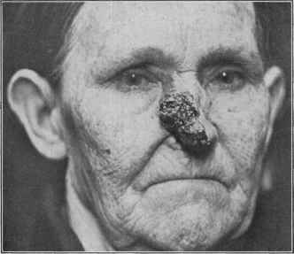
Fig. 222.—Epithelioma of papillomatous type in a woman aged sixty-three.
lowed by recurrence and finally death. Epithelioma of the back of the
hand is usually responsive to proper measures, but the prognosis is not
so favorable as with the ordinary face cases, and axillary gland involve
ment sometimes presents early. Epithelioma of the genitalia is always
of serious import, although prompt action in the beginning disease is
commonly successful.
Treatment.—The object to be kept in view in the treatment of
the disease is the thorough destruction or removal of the epitheliomatous
tissue. For this purpose operative measures are preferred by the sur
geon,1 while the specialist in dermatologic practice usually favors the
caustic plans. To a great extent, I believe this difference of opinion to
1 Bloodgood, “The Surgical Treatment of Malignant Growths,” Jour. Amer. Med.
Assoc, 1910, lv, p. 1615 (based on malignant pigmented moles 65, Sarcoma of the derma
45, and epithelial tumors of the skin and mucous membranes 812, and benign moles 75).
CARCINOMA CUTIS
885
be due to the fact that those cases coming under our care are relatively
superficial and slight, circumscribed, and slow, whereas those coming
to the knowledge of the surgeon are for the most part of a more serious,
malignant, and extensive nature. The former do well and probably
better with caustic methods, the latter with surgical measures. With
well-defined, sharply circumscribed epitheliomata, especially if of the
deep-seated and papillomatous varieties, surgical removal is to be given
the preference, particularly if the lesion is seated upon parts of loose
and soft texture, where excision can be followed by approximation of the
edges and thus leave but a linear scar. When the neighboring glands
are implicated, these, too, should be extirpated. Such cases, however,
naturally belong to the domain of surgery. A combination of the surgical
plan of thoroughly curetting the area and then following with caustic,
such as the application of a 50 per cent, solution of zinc chlorid or 60
per cent, acid nitrate of mercury solution,1 or momentary cauterization
with caustic potash or with several days’ use of a strong pyrogallol salve,
to be referred to later, constitutes a successful method, applicable in
many instances. This latter plan is to be commended for epitheliomata
of moderate size and infiltration. For those instances, however, in which
the slightest mention of operative procedure is met with opposition
or withdrawal, the caustic plans of treatment can be resorted to; and for
the superficial skin cancers, those, as a rule, without glandular involve
ment, and which often come to the specialist in dermatology, I am con
vinced, from considerable experience, that the method is not only a
practicable one, but usually permanently successful.2 The effect of the
caustic seems to extend beyond the escharotic action produced.
The favorite caustics are pyrogallol, zinc chlorid, caustic potash,
and arsenious acid. After their use the subsequent treatment consists,
when possible, of the continuous application of poultices until the slough
comes away, and then a mild healing ointment of 1 to 2 per cent, pyrogal-
lol salve, one of equal parts of mercurial plaster and petrolatum, or of
zinc oxid. When poultices are not practicable, the healing ointment
can be immediately applied after the cauterization; in such instances,
however, the slough comes away much more slowly. The part is washed
once or twice daily, and oftener if there is much discharge. Pyrogallol
is applied in the form of a salve of 25 to 40 per cent, strength, in the man
ner described under Lupus vulgaris, and continued for from one to two
or three weeks, according to the character of the growth and the rapidity
of action. A good formula consists of the following:
R. Pyrogallol, 3iiss-iij (10.-12.);
Ac. salicylici, gr. xxv-1 (1.65-3.33);
Cerat. simp., 3j-ij (4._8.);
Petroláti, q. s. ad 3j (32.).
1 Sherwell, Jour. Cutan. Dis., 1910, p. 487 (with a number of excellent case photo
graphs), has had remarkable success with the method of treatment-curetting and
supplementary cauterization with the acid nitrate of mercury solution.
2 See papers on the caustic treatment by Robinson, Internat. Jour. Surg., 1892, p.
179, and 1893, p. 164, and New York Med. Rec., Mar. 31, 1900; Gottheil, “The Treat
ment of Skin Cancers,” New York, 1899; Stelwagon, Jour. Amer. Med. Assoc, Dec 15,
1900; and Heidingsfeld, ibid., July 13, 1901; Van Harlingen, Jour. Cutan. Dis., 1906,
p. 345, strongly commends the caustic potash treatment.
886
NEW GROWTHS
The amount of cerate depends upon the season, whether warm or cold.
The formula also mentioned under Lupus is equally available, and is
sometimes to be preferred on account of greater adhesiveness. Pyrogal-
lol is a relatively painless application, and has its chief field in superficial
epitheliomata in old people who cannot bear well the stronger caustics;
and also is of great value as a supplementary caustic to curetting, as
already remarked. It is, however, a weak caustic and has a limited
field. Like arsenic, it generally spares the healthy tissue.
Zinc chlorid is a caustic formerly much in vogue for its destructive
action. It is painful and destroys morbid and healthy tissue alike.
Its action is peculiar in that it seems to mummify the tissue, and there
fore is especially applicable to epitheliomata situated over blood-vessels.
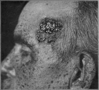
Fig. 223.—Epithelioma of papillomatous type; showing also old-age changes—freckle-
like pigmentation and scurfy patches.
It is most commonly applied as Bougard’s paste, but for satisfactory
compounding it requires ordinarily somewhat more water than given,
although it is advisable to exceed the quantity in the formula as little
as possible, so that the paste may be like stiff dough, and then to add at
the time of application a siifncient quantity of a saturated solution of
cocain hydrochlorate to bring it up to working consistence. The for
mula is as follows: R. Farinæ trit. (wheat flour), pulv. amyli, ää 3ss
(16.); pulv. ac arseniosi, gr. iv (0.26); pulv. hydrarg. sulph. rub., pulv.
ammonii chlorid., ää gr. xx (1.33); pulv. hydrarg. chlorid. corr., gr. ij
(0.135); zinci chlorid. cryst., 3iv (16.); aquæ fervid., 3j (32.). The first
six ingredients are thoroughly mixed, the zinc chlorid dissolved in the
water, and the two mixtures rubbed up together secundum artem.
CARCINOMA CUTIS 887
This is spread on any suitable material and applied to the growth, ex
tending slightly beyond the border. The depth destroyed is usually
one or two times the thickness of the layer of paste. It takes about
twenty-four to forty-eight hours for sufficient destruction; it is some
times necessary to remove the mummified mass by paring it away
and reapplying a fresh plaster. There is considerable inflamma
tory swelling. In superficial lesions rarely more than one application
is necessary. The separation of the slough requires from five to twenty
days.
Caustic potash is a powerful caustic and must always be used with
care. It is rapid in its action, one thorough application usually sufficing
to destroy the entire growth. It has its special field in small and be
ginning cutaneous skin cancers, and in those in which time is important
and in which the patient can remain under observation only a short
period. The stick should be employed, or the strongest possible solution;
the former is preferable. If the surface of the growth is crusted, this
should be removed, and the parts outside of the diseased area protected
with a layer of vaselin, after which the caustic is applied. If the lesion
is small and superficial, the desired effect is usually sufficiently attained
in a minute or two, and then further action is to be prevented by the
application of dilute acetic acid or vinegar. This caustic is painful, but
only at the time of application.
Arsenic is undoubtedly the best caustic to employ in many of these
cases. It certainly has, relatively speaking at least, an elective action,
ordinarily sparing healthy tissue, so that it is especially applicable in
places where unnecessary destruction and disfigurement are to be avoided,
as particularly about the nose and in the neighborhood of the eyelids. It
should not be applied to a large surface, not larger than a square inch.
Marsden, Robinson, Gottheil, and others, including myself, who have
employed it frequently, have never seen dangerous absorption from its
cautious use. It may be employed in several strengths, according to the
case and the effect required. Marsden advised a paste (Marsden’s
paste) made of two parts of arsenious acid and one of mucilage of acacia.
Robinson recommends two strengths, one of equal parts of arsenious
acid and powdered acacia, and one of two parts of the arsenic and one
of the acacia, using sufficient water at the time of application to make into
a paste of the consistence of stiff butter. I have employed it, in most
cases, in about equal proportions, in small and somewhat deep-seated
lesions, using two or three parts of arsenious acid to one of acacia, making
up into a paste with a saturated solution of cocain hydrochlorate. It
requires from twelve to thirty-six hours for sufficient action, producing
a good deal of inflammatory swelling and edema; occasionally a second
application is necessary. It is painful, but many patients prefer it to
surgical operation. It is the principal ingredient in most of the quack
cancer plasters. The slough separates slowly.
Recently carbon-dioxid snow (q. v.) has been extolled for its favorable
cauterizing action in the small superficial and beginning rodent ulcer
types.
Electrolysis has also had occasional advocates for certain mild or
888 NEW GROWTHS
slight cases—with the needle in small lesions or recurring small tubercles
in the scar after other treatment; and with the small metallic plate, as
in lupus vulgaris, in small flat growths. If used with a current of 5 to
20 milliampères, it is capable of active destruction.
The treatment of epithelioma by the Finsen concentrated chemic
light method (see Lupus vulgaris) has been favorably reported upon
by some writers, among whom the latest, Bie,1 who had, in 16 cases,
a good proportion of satisfactory results. Finsen regards it as most
favorable in those cases which are superficial and well demarcated.
According to these observers, about 30 exposures, of about one hour
each, are required, the growth gradually shrinking and healing.
The plan of treatment most in vogue at the present moment is that
by the x-ray. The experience of Williams, Pusey, Pfahler, and many
others, including myself, attests its favorable and sometimes curative
action. It is usually slow, and on this and other accounts is not, as a
rule, to be advised as the sole measure of treatment; in superficial forms,
especially when involving the neighborhood of the eye, it often acts well
and comparatively quickly. Its special field of usefulness is, in my
opinion, as a supplementary measure to those methods already practised
and described above. To secure a result it must often be pushed to the
point of producing a mild erythema or even moderate dermatitis. Ex
posures are made every one to three days, at a distance of 3 to 15 inches,
and of six to twenty minutes’ duration, depending upon the effect; and
in superficial cases or those of but slight or moderate depth with a soft
to medium tube. It is not a good plan, however, to use a tube of the
same degree of vacuum throughout; otherwise some possible deeper-
lying morbid tissue may escape its favorable action. I have found it a
good plan to begin the séance with a vacuum equal to about a 1/2-inch
spark, and then to do away with the regulator; the vacuum slightly rises,
and the whole depth of tissue gets its share of the full effect of the rays.2
The surrounding parts are to be protected by thin lead foil. The first
effects consist of a drying-up of the secretion and a gradual shrinking
of the ulcerated area. Some cases are much more responsive than others,
and a few fail to show any marked change. In some cases favorable
influence and cure in cancerous growths follows the application of
radium,3 and in a few instances under my own care it has been of helpful
service.
From time to time various remedies have been suggested for con
stitutional administration as having a favorable action in certain in
stances; arsenic has had the most frequent mention, but, excepting a
few observers, it has but little support. Sherwell4 is convinced of its
value, and makes its administration a routine practice, along with proper
1 Bie, Dermatolog. Zeitschr., Aug., 1900, p. 630—abs. in Brit. Jour. Derm., 1900,
p. 376.
2 The details on x-ray treatment are more fully given under General Remarks on
Treatment, in the first part of the volume.
3 Abbe, “Radium in Surgery,” Jour. Amer. Med. Assoc, 1906, vol. xlvii, p. 183;
Wickham and Degrais, “Radiumthérapie,” Paris, 1909.
4 Sherwell, “The Use of Arsenic, etc, in Cancerous and Other Neoplasms,” Med.
Record, April 28, 1900.
XERODERMA PIGMENTOSUM 889
local measures; Lassar1 has also reported favorable effects, and Pusey2
thinks it may have an inhibitory action.
But first, if you want to come back to this web site again, just add it to your bookmarks or favorites now! Then you'll find it easy!
Also, please consider sharing our helpful website with your online friends.
BELOW ARE OUR OTHER HEALTH WEB SITES: |
Copyright © 2000-present Donald Urquhart. All Rights Reserved. All universal rights reserved. Designated trademarks and brands are the property of their respective owners. Use of this Web site constitutes acceptance of our legal disclaimer. | Contact Us | Privacy Policy | About Us |