| MEDICAL INTRO |
| BOOKS ON OLD MEDICAL TREATMENTS AND REMEDIES |
THE PRACTICAL |
ALCOHOL AND THE HUMAN BODY In fact alcohol was known to be a poison, and considered quite dangerous. Something modern medicine now agrees with. This was known circa 1907. A very impressive scientific book on the subject. |
DISEASES OF THE SKIN is a massive book on skin diseases from 1914. Don't be feint hearted though, it's loaded with photos that I found disturbing. |
FIBROMA
Synonyms.—Molluscum simplex; Molluscum fibrosum; Fibroma molluscum;
Molluscum pendulum; Molluscum non-contagiosum; Fr., Fibrome; Nævus mollus-
coide; Molluscum vrai; Ger., Fibrom.
Definition.—Fibroma is a connective-tissue new growth, appear
ing as one or more sessile or pedunculated, pea- to egg-sized or larger,
soft or firm, rounded, sometimes flattened, painless tumors, seated be
neath and in the skin.
Symptoms.—The tumors appearing in this disease show varia
tions as to size, shape, and numbers. There may be but a single growth
or they may be numerous. Occurring as a single tumor, which is the
more common, it is usually more or less pedunculated, and, when
reaching any great size,—and it quite frequently attains considerable
dimensions,—it becomes pendulous (fibroma pendulum). In the mul
tiple cases the growths may be somewhat scanty in number, or may exist
694 NEW GROWTHS
in great profusion, as in the instances observed by Octerlony,1 Hewson,
and others; in extreme examples they may be present in such abundance
as to crowd the surface, as in the case reported by Dunn.2 In these
extensive cases the growths vary from a pea to an egg or larger, and may
be almost all more or less rounded and sessile, although usually some show
a trifling or moderate tendency to narrowing at the base, giving the tu
mors a pear shape, and such, when the narrowing is at all marked and
the growths moderately large, are generally slightly pendulous. Others
may be sausage shaped, and exceptionally show a tendency to lobulation.
In other cases the tumors will be, for the most part, as just described;
but one or several extremely large pedunculated growths (fibroma pen
dulum) will be present, with a comparative small pedicle and a variously
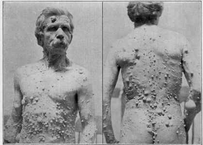
Fig. 156.—Fibroma (front and back view of the same patient) (courtesy of Dr. Addinell
Hewson).
sized, pear-shaped, often somewhat flattened, pendulous mass, which
hangs down and often covers up some of the smaller tumors. In these
general cases the upper part of the back seems to be a favorite region
for the pendulous growth, as in Tappey‘s and Iurkewicz‘s3 patients.
The smallest tumors project but slightly, in some instances appearing
1 Octerlony, Arch. Derm., 1875, P- 300, having 2333 growths (with illustrations);
Wigglesworth, in the same journal for 1876, p. 193, also records a similar case (with
illustration), having 1193 tumors; Hashimoto, Sei-I-Kwai Med. Jour., Dec, 1888, p.
197 (with illustration), described a case with 4503 growths; Pooley, Jour. Cutan. Dis.,
1894, p. 117, has also published an extensive case (with illustrations).
2 Dunn, Med. Press and Circular, 1890, p. 623 (with good illustrations); a plate of
this remarkable case, credited to Hutchinson, will also be found in Morrow‘s System,
vol. iii (Dermatology), op. p. 478.
3Tappey, Jour. Cutan. Dis., 1889, p. 179 (with illustration); Iurkewicz, Meditzin-
koië Obozrenië, No. 21, 1891, p. 738 (with drawing)—abs. in Brit. Jour. Derm., 1891, p.
367.
FIBROMA
695
to be practically subcutaneous, although in other cases they are inti
mately associated with the skin proper and are more elevated. In the
moderate and larger sized growths the elevation is conspicuous, and when
narrowing of the base is present, they are essentially situated wholly
above the surrounding level.
The skin over the tumors is generally normal, but it may be tense or
lax, and of a natural pinkish or reddish color. The reddish color is
generally seen in those growths which develop rapidly, the slowly grow
ing tumors—the usual course—remaining more or less normally colored.
In some tumors, more especially those of larger size, the openings of the
sebaceous glands are enlarged and hypertrophied, and sometimes con
tain blocked-up secretion or plugs. In other instances, usually in those
lesions in which the skin is tense and distended, the follicles may be
atrophied and the integument somewhat thinned. To the touch they usu
ally feel soft or doughy and slightly elastic, and are painless. They do
not undergo destructive change, although with the heavy, pendulous
formations, as a result of weight or pressure, surface abrasion and ulcera-
tion may occur; and when crowded together, owing to their number,
size, and location, as a result of interference with motion or by accidental
injury, the larger growths may occasionally become inflamed, and ex
ceptionally undergo ulceration and even become gangrenous. In some
of the rapidly developing tumors the skin, which becomes red and vas
cular, may later become excoriated and even ulcerated. Gangrenous de
struction also occasionally occurs in the growths with extremely thin
pedicle. Ordinarily, however, such accidents do not occur, and except
for the disfigurement and discomfort of their presence, they give rise to
no serious condition. In the course of time, but usually slowly, some
lesions continue to increase in size, new ones may arise, while others,
having obtained variable dimensions from small to large, remain more or
less stationary, so that there are usually to be seen, in a given case,
tumors of all sizes from that scarcely larger than a pin-head or small
pea to that of considerable proportions; the latter, especially the large
pendulous tumors, sometimes reaching huge size, and weighing many
pounds. The greatest size and weight are observed in the single fibroma,.
although in the multiple cases sometimes one or two tumors also attain
enormous development. In the average case there seems to be a steady,
usually slow, increase in the number of the growths, although after a time
the malady is apt to remain stationary; exceptionally, however, there
are seen to be periods of active increase, and this has been more especially
noticed in women and in connection with pregnancy (Hirst1). Indeed,
there is a peculiar small type growth observed occasionally in pregnant
women, presenting about the fourth to the sixth month of pregnancy,
gradually increasing, as a rule, in numbers (rarely exceeding 50) up to
full term, and then slowly, in the course of a few months disappearing
(Brickner2); they are usually only seen about the neck, breast and sub-
mammary region.
1 Hirst, “A Note on the Etiological Influence of Pregnancy upon Molluscum Fibro-
sum,” Amer. Jour, of Obstetrics, 1911, lxiii, p. 256.
2 Brickner, “Fibroma Molluscum Gravidarum,” Amer. Jour, of Obstetrics, 1906,
liii. p. 191 (with histologic report by Pollitzer).
696
NEW GROWTHS
Occasionally the tumor growths are ill defined, consisting of irregular
and nodular, confluent, wrinkled, and fold-like masses, and when such
formations are numerous and of gigantic size, they give the patient in
regions an elephantine appearance—the extreme development, which
seems really a combination of fibroma, elephantiasis, and dermatolysis,
giving rise to the appellation “elephant man,”1 In this instance there
were also some exostoses. Occasionally a growth, usually those of mod
erate size, undergoes partial involution or absorption of the interior por
tion, and hangs like a flaccid, partly filled pouch or sac This absorption
exceptionally takes place in the large pedunculated or sessile growths,
and when more or less complete, results in a soft mass of pendant, vari
ously hypertrophied skin—dermatolysis (q. v.). This same change is
also at times noted in the small pea-sized, isolated fibromata, and fleshy
moles, soft warts, or solid, warty-looking growths, in the skin of those
advancing in years (see Atrophia senilis), which are often pedunculated,
and which result in small, pendulous sacs; to these the name of fibroma
simplex, or acrochordon, is sometimes given, although the term fibroma,
when employed, usually refers to the more pretentious growths, which
have been described, but which, as remarked, may undergo similar invo-
lutionary changes.2 Occasionally, in a pea- to cherry-sized growth, more
especially the smaller, when such absorption or involutionary change
takes place, there remains a slight projection, seemingly hollow and read
ily compressible, and sometimes of a bluish tinge.
In some instances neurofibromata have coexisted, as in the cases
recorded by Atkinson,3 von Recklinghausen,4 Payne,5 Brigidi,6 Briquet,7
and others.8 In some cases, as Recklinghausen and some others believe,
the lesions are doubtless all neurofibromata (Recklinghausen‘s disease,
neurofibromatosis). Other lesions sometimes associated are brownish,
pigmentary stains, sometimes freckle-like, small or large areas, and
occasionally more or less diffused discoloration. While, as Wickham9
1 Editorial report of an extreme example in Brit. Med. Jour., 1886, ii, p. 1188 (with
illustrations); an abbreviated account, with illustrations, also in Jour. Cutan. Dis., 1887,
p. no.
2 See Taylor‘s paper, “Molluscum fibrosum and its Relation to Acrochordon and
other Cutaneous Outshoots,“ Jour. Cutan. Dis., 1887, p. 41; and to “Keloid,” ibid., p. 161.
3 Atkinson, New York Med. Jour., 1875, vol. xxii, p. 601 (2 cases in family).
4 Von Recklinghausen, Ueber die multiplen Fibrome der Haut, und ihre Beziehung
zu den multiplen Neuromen, Berlin, 1882 (a résumé of fibroma cases; 5 plates, 2 of case
illustrations and 3 histologic). 5 Payne, Brit. Med. Jour., 1889, i, p. 592.
6 Brigidi, Monatshefte, 1894, vol. xix, pp. 190 and 237 (with histologic cuts and
bibliography). 7 Briquet, Jour. med. cutan., 1898, p. 219 (with bibliography).
8 Whitfield, Lancet, Oct. 31,1903, p. 1230 (newly formed nerve-fibers were found in
the growth); Krzystalowicz, Monatshefte, 1903, vol. xxxvi, p. 421 (case report, histologic
review, and bibliography to date); Piolett, Hospital Gazette, 1902, No. 137, brief ab
stract in Jour. Cutan. Dis., 1905, p. 363 (with more than 600 tumors), Benaky, Annales,
1905, p. 977; Merk, Archiv, 1905, vol. lxxiii, p. 139.
9 Wickham, Paris letter, Brit. Jour. Derm., 1890, p. 151; Parkes-Weber, Brit. Jour.
Derm., 1909, p. 49, reviewing the subject, calls attention to the fact that cases of Reck-
linghausen‘s disease occur in which decided pigmentation of the skin is developed long
before neurofibromata of nerve-trunks or molluscum tumors of the skin are observed;
Ravogli, “Fibroma Molluscum, or Universal Neurofibromatòsis,” Jour. Cutan. Dis.,
Feb., 1911 (records a case; illustrations, review of the subject in general, with good
bibliography); Friedlander, “Multiple Neurofibromata” Jour. Cutan. Dis., 1910, p.
497, reports a case and gives review, based on 262 cases reported in the literature
(good bibliography).
FIBROMA
697
states, some authors touch upon this feature, present in many cases, by
others it is entirely ignored. In Wickham‘s 8 generalized cases such
pigmentary conditions were present in all; and in addition there were
small, violaceous, compressible prominences, already noted, but which
Wickham apparently considers arise as such, and not necessarily as a
result of involutionary changes.
Any part of the surface may be the seat of fibromata, but, as a rule,
the tumors are most numerous and largest on the trunk, both front
and back. The scalp1 and other parts of the head are also favorite
localities, and the extremities usually show the smallest number. The
palms and soles are rarely invaded, and, when so, the growths are small
and flattened. In some instances they have also been found on the mu
cous membranes, as on the lips, gums, hard palate, and tongue.
Another form of fibroma, called hard fibroma,2 or desrnoids, in con
tradistinction to that which is ordinarily met with and just described
(sometimes called soft fibroma) is in many respects similar to the more
solid small growths already referred to as fibroma simplex. They
are rarely larger than a pea, occur usually singly, or as several
scattered solid growths, covered by normal skin; are sharply defined,
round or oval, smooth and compact, and movable. Their appearance
is insidious and their growth slow, and they may appear at any age, and
are even present, in some instances, at birth.
Etiology.—The affection is not a common one in our own
country, England, or the Continent, but is, according to Hashimoto,
quite frequent in the eastern countries. Its etiology is obscure, although
it is known, from the observations of Virchow,3 Konigsdörf,4 Octerlony,5
Atkinson,6 and others, to have occurred in several successive generations,
or sometimes in more than one member of the same family. Heredity
or family tendency must, therefore, be considered a factor. It occurs
in both sexes, in all nationalities, and usually begins in childhood and fre
quently in early infancy; in some instances it is congenital (Hahn, Tap-
pey, Hallopeau, and others).7 In early life the lesions are, however,
small and relatively scanty, and the increase in size and number takes
place very slowly, as a rule, not developing to any extent until much
later. In the cases of single fibroma its appearance is, as a rule, later in
life. The subjects of the malady, as Hebra pointed out, and also shown
in those of Pooley, Pringle,8 Iurkewicz, and many others, are often of
1W. G. Smith, Brit. Jour. Derm., 1896, p. 115, describes and illustrates a case of
extensive, somewhat lobulated fibroma, seated upon the scalp, with no tumors else
where.
2 Synonymous with Unna‘s “ Fibroma simplex.” Recently, under the name of
“Noduli cutanei,” Arning and Lewandowsky (Archiv, 1911, cx, p. .3) reported a series
of cases (20 occurring among 5000 patients) which histologic examinations indicated
to be the same formation; evidently this form is not so uncommon as thought, but
from their benign and painless character often overlooked.
3 Virchow, Virchow‘s Archiv, 1847, vol. i, p. 226 (according to the patient‘s state
ment his grandfather, father, brother, and sister had the same disease).
4 Königsdorf, “Ein Fall von Fibroma Molluscum Multiplex,” Dissertation, Würz-
burg, 1889 (quoted by Jarisch).
5 Octerlony, loc. cit. (a brother also had it). 6 Atkinson, loc. cit.
7 Hahn, “Beiträge zur Casuistik des Fibroma Molluscum,” Dissertation, Würzburg,
1888; Hallopeau, Annales, 1889, p. 707 (case demonstration and histologic examination).
8 G. L. K. Pringle, Edinburgh Med. Jour., 1900, vol. xlix, p. 260 (with plate).
698
NEW GROWTHS
weak physical and of defective mental development, but while so in the
larger number of cases, it by no means obtains in all. Moreover, it
is not improbable, as Hutchinson1 suggests, that the mental apathy is
the indirect result of the gross disfigurement, the patient holding himself
aloof and shunning his fellows. Traumatism is thought a possible
determining factor in their production, or, more probably, only in in
fluencing their location, more especially in the single fibroma developing
later in life. Schwimmer, Taylor, and Recklinghausen, as well as a
few others, have noted this, the last calling attention to the fact that those
parts of the body most subject to friction, pressure, etc, usually show
the most numerous growths. In some instances in women pregnancy
seems, directly or indirectly, of some etiologic influence (Brickner,
Hirst).
Pathology.—According to the investigations of Rokitansky,
Virchow, Neumann, Sangster, Duhring, Crocker, and others, the growth
is due to a hyperplasia of the connective tissue, although there is not
the same unanimity as to its exact starting-point, whether from the con
nective tissue of the corium, of the framework of the fat-globules, or
of the walls of the hair-follicles and sebaceous glands. As to what gives
rise to this hyperplasia is unknown. Recklinghausen, from his investi
gations of multiple fibromata, believes that they are really neurofibro-
mata, and that they are formed primarily by proliferation of the con
nective-tissue sheaths of the nerves, and subsequently added to by pro
liferation of the same tissue of sweat-glands, sebaceous glands, and blood-
vessel sheaths. The admixture of neurofibromata in some cases is
generally recognized, but that fibromata, as commonly met with, are
all of the same origin or nature is negatived by the collective investiga
tions of others. Both Pringle2 and Anderson3 have also called attention
to the fact that there is sometimes an association of fibromata with ade
noma sebaceum, and it is not at all impossible, therefore, as these several
gentlemen suggest, that certain tumors of different origin and character,
which are sometimes found together, may have some common pathologic
relationship. Crocker suggests that the growth may be due to obstruc
tion of the superficial lymphatics, and that this, as well as other, anatomic
analogies bring it into pathologic relationship with elephantiasis.
As the beginning lesions grow and extend the skin is pushed upward,
and they finally project as simple or lobulated, sessile or pendent tumors;
they are adherent to the skin only at their base, and may thus be easily
enucleated (Heitzmann). Crocker states that a sebaceous gland or hair-
follicle forms the center in many of the small tumors, while these struc
tures in the larger or older growths have undergone atrophy or disap
peared. According to Taylor, in their very earliest stage the tumor con
sists of a gelatinous structure, which, under the microscope, is found
composed of a succulent, edematous, wavy connective tissue with many
cells, while in the older growths the fibers are firm and not edematous,
1 Hutchinson, “Molluscum fibrosum,” Rare Diseases of the Skin, p. 205.
2 Pringle, “Case of Congenital Adenoma Sebaceum,” Brit. Jour. Derm., 1890, p. 1.
3 Anderson, “A Case of Adenoma Sebaceum Intermingled with Mollusca Fibrosa,”
ibid., 1895, p. 316.
FIBROMA 699
and the cells are less numerous. On cutting through a well-formed
tumor of some duration, quoting chiefly from Crocker and Heitzmann,
it is found to consist of a white, fibrous mass, inclosed in a dense con
nective-tissue capsule, with the central portion soft and pulpy, and from
which a small quantity of clear yellow fluid can be pressed out. The
fibrous tissue is firmest and most developed at the base, the fibers becom
ing less firm and softer as the interior is approached. Connective-tissue
cells with large nuclei are found between the fibers, being most numerous
in the gelatinous central portion. The vascular supply consists of large
afferent and efferent vessels, readily demonstrable at the base, and which
spread peripherally, terminating in fine capillaries. The epidermis
remains unchanged, although the sebaceous gland-ducts are sometimes
hypertrophied, patulous, and plugged with comedones.
Diagnosis.—In a large single and pedunculated fibroma, and in
cases of multiple, scattered, variously sized growths, most of which are
sessile, and possibly a few with a narrowed neck or pedicle, a correct
diagnosis is a matter of no difficulty. Confusion is most likely to occur
with multiple lipoma, but in this latter they are commonly lobular,
somewhat flattened, rarely present in numbers, and never pedunculated.
From multiple neuromata they are to be distinguished by the absence
of pain, as well as usually by their more general distribution. A mistake
has sometimes been made with molluscum contagiosum, but the growths
of the latter are much smaller, rarely numerous, most commonly seated
about the face, especially about the eyelids, and, moreover, are super
ficial, have a central depression or aperture, and are covered by skin,
which is usually thin, stretched, and which has a semitranslucent appear
ance. There might also be a possibility of confusing fibroma with the
early tumor stage of granuloma fungoides, but the usual preceding and
accompanying eczematoid symptoms of the latter, as well as the tend
ency, in some growths, toward the formation of fungoidal ulcerating
masses, and the late development and sometimes capricious behavior
of the tumors,—appearing and disappearing,—are wholly different from
the features of fibroma. The soft and warty moles, sometimes congenital
and sometimes developing later in life, can scarcely be confounded with
fibroma, as commonly understood, although such growths, usually small
and few in number, and generally more or less pigmented, are to a great
extent to be placed in the same category. In the similar lesions observed
in old people about the face and back, the surface is, as a rule, dark col
ored, often warty, and frequently covered with a greasy scale or crust.
It is scarcely likely that fibromata could be confused with the nodules
of leprosy, sebaceous cysts, or gummata.
Prognosis.—The outlook, so far as life is concerned, is always favor
able, but as to the growths themselves they are persistent, and usually
add gradually to their size and also increase in numbers. While not
therefore, involving the general health, still, by their presence, they often
give rise to inconvenience and discomfort by interfering with freedom of
motion and through accidental injury, besides being the source of mental
worry, which sometimes leads to an apathetic or neurasthenic condition.
Exceptionally, in some growths, a tendency to spontaneous involution
7oo
NEW GROWTHS
is exhibited. In single fibroma, and also in moderate multiple cases,
there may be relief or comparative freedom brought about through
operative procedures. Unfortunately, the malady is not influenced by
constitutional treatment, although one instance of great improvement,
to be later referred to, has been recorded.
Treatment.—Surgical measures alone are of any reliance, and
the method, whether by ligation, écraseur, galvanocautery, or excision,
depends upon the size and character of the growths. Large pendulous
tumors can always be removed readily, and with great relief to the pa
tient. The smaller tumors also admit of removal if not too numerous
and if done a few at a time. The huge, flabby growths, approaching the
nature of dermatolysis, have also been excised with success. Whatever
the method, the tumor should be thoroughly extirpated or a regröwth
is probable. Electrolysis has proved serviceable for the small tumors.
In multiple cases, in view of the favorable influence from the long-
continued administration of arsenic exceptionally observed in other
multiple tumor growths, this drug might also be tried in fibromata,
more especially so now in view of the favorable influence apparently
exerted in a case under Whitehouse's1 care.
But first, if you want to come back to this web site again, just add it to your bookmarks or favorites now! Then you'll find it easy!
Also, please consider sharing our helpful website with your online friends.
BELOW ARE OUR OTHER HEALTH WEB SITES: |
Copyright © 2000-present Donald Urquhart. All Rights Reserved. All universal rights reserved. Designated trademarks and brands are the property of their respective owners. Use of this Web site constitutes acceptance of our legal disclaimer. | Contact Us | Privacy Policy | About Us |