| MEDICAL INTRO |
| BOOKS ON OLD MEDICAL TREATMENTS AND REMEDIES |
THE PRACTICAL |
ALCOHOL AND THE HUMAN BODY In fact alcohol was known to be a poison, and considered quite dangerous. Something modern medicine now agrees with. This was known circa 1907. A very impressive scientific book on the subject. |
DISEASES OF THE SKIN is a massive book on skin diseases from 1914. Don't be feint hearted though, it's loaded with photos that I found disturbing. |
KELOID
Synonyms.—Cheloid; Alibert‘s keloid; Kelis; Kelos; Fr., Chéloide; Kéloide.
Definition.—Keloid is a fibrocellular new growth of the corium
appearing as one or several variously sized, irregularly shaped, elevated,
smooth, firm, pinkish, or pale-reddish cicatriform lesions.
Symptoms.—The growth begins as a small, hard, elevated,
occasionally somewhat deeply imbedded, pinkish or reddish tubercle
or nodule, increasing gradually in size. Usually months or years elapse
before the tumor reaches conspicuous dimensions. In fact, not infre
quently, and more especially in multiple cases, the growth increases
but slowly, and, after attaining small proportions, sometimes scarcely
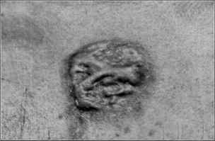
Fig. 149.—Keloid; over sternum.
greater than a large pea or bean, remains stationary more or less in
definitely or permanently. These small growths are of a pinkish-
white or reddish color, firmly seated in the corium, distinctly elevated,
and usually smooth and glossy, with a rounded or somewhat flattened
top, and with almost perpendicular or slightly sloping sides. It is hard,
and the surface may show, on close inspection, one or two capillaries.
Ordinarily, however, and particularly in the single growth, it gradually
increases in size, spreading laterally by an invasion of the surrounding
skin, and frequently extending upward as well, sometimes finally reach
ing considerable elevation. Very commonly the border extends out
ward in the shape of several or more claw-like projections; to this feature
is owing the name keloid. The process may go on slowly or somewhat
rapidly, and in extreme cases a large area may be involved and enormous
proportions reached.
In average cases, when developed, the growth is observed to be one,
1 Varney, Internat. Jour. Surg., Oct., 1903, p. 309.
KELOID
637
several, or more inches in diameter, is sharply defined, elevated, hard,
rounded or oval, fungoid or crab-shaped, and firmly implanted in the
skin, and having a scar-like aspect. It is of a pinkish, pearl white, or
reddish color, commonly devoid of hair, with no tendency to scaliness,
and with usually several vessels coursing over it. In some instances
it is elongated and ovalish. The surface, which is generally shiny and
glistening, with the epidermis having a stretched and tense appearance,
is flattened or irregularly rounded, or with slight nodular projections;
often the central part is slightly lower than the main and peripheral
portions. Sometimes, instead of the colors just named, it is of slight or
distinctly purplish hue; and occasionally, in place of the rounded, ovalish,
lozenge or crab-shaped growth, the formation is exceedingly irregular,
with prolongations which may extend to a considerable distance, and
from which also may go claw-shaped extensions; exceptionally it is
streak- or band-like. In general the height is about \ to \ inch, although
in the enormous keloids it may reach several inches or more, the whole
growth assuming large, tumor-like proportions.
While in many instances there are no subjective symptoms,—which,
in fact, may be said to be the rule,—in others itching or tenderness is
complained of, and occasionally it is spontaneously painful. The most
frequent situation for keloid is over the sternum, although other parts
of the upper trunk are often the site of the growth; it may also appear
on the face, ears, neck, and extremities. Commonly but one or two
lesions are present, but there may be several or more up to a considerable
number, as in the instances observed by Wilson,1 Schwimmer,2 De
Amicis,3 Goodhart,4 Smith,5 Hardaway,6 and others. In those cases
following smallpox the lesions are usually numerous, though, as a rule,
small. In some of the instances of multiple keloid the growths are more
or less symmetrically arranged, as in the cases of De Amicis and Smith
just referred to, and also in an instance observed by Vidal.7
Etiology.—The cause of keloid is not known. It has been the
custom to divide these growth into two varieties—those that arise
at the site of burns, cuts, acne, syphilis scars, etc, designated scar keloid,
cicatricial keloid, false keloid, spurious keloid, secondary keloid, and those
that are believed to originate in normal and uninjured skin, as idiopathic
keloid, primary keloid, spontaneous keloid, true keloid. In later years
there has, however, been less and less tendency to make these two
1 E. Wilson, Diseases of the Skin, 1867, p. 381 (39 growths—30 on the breast, 9 on
back), cited by Schwimmer.
2 Schwimmer, “Die multiple Keloid,” Archiv, 1880, p. 225 (105 growths, more or
less general, with review of the subject and principal references to date; histologic
report of this case by Babesiu, p. 237).
3 De Amicis, “Chéloide spontanée multiple,” Trans. Internat. Dermatolog. Cong.,
Paris, 1889, p. 93 (318 growths, symmetrically on the scapulohumeral regions and arms;
3 colored plates).
4 Goodhart, London Clin. Soc‘y Trans., 1880, vol. xiii, p. 51 (development from
smallpox scars—numerous and quite pronounced, with colored plate of face).
5 W. G. Smith, Brit. Jour. Derm., 1889, p. 157 (numerous, but number not stated,
more or less general, and, upon the whole, a decided tendency to symmetry).
6 Hardaway, Manual of Skin Diseases, second edit., case illustration op. p. 287
(negro—with numerous lesions on trunk and arms).
7 Vidal, Trans. Internat. Dermatolog. Cong., Paris, 1889, p. 103 (12 growths, sym
metric over shoulders and nape of the neck).
638 NEW GROWTHS
divisions, and the doubt of a keloid arising without a slight traumatism
is pretty generally entertained. When we consider that the injury which
often seems to start the pathologic process may be extremely slight,
such as scratching, insect-bites, slight pricks, and the like, .it can readily
be seen how such could be easily overlooked or actually be so insignificant
as to go unrecognized, and thus give rise to the assumption that the
keloid was spontaneous. Even the more or less general cases, such as
those of De Amicis, Schwimmer, and others, which are apparently spon
taneous, and which are usually quoted as convincing examples of this
variety, could be readily explained upon the assumption of such trifling
abrasions or injuries as just noted. There is, therefore, in my judgment,
considerable ground for Unna‘s1 opinion that the most frequent site for
the so-called spontaneous keloid growth, over the sternum and about
the breast, is due to the irritation and scratching invoked by dermatitis
seborrhoica, so common in this region. Crocker suggests that possibly
the frequency in this region “may be accounted for, in women, by the
pressure and friction of the stays, and, in men, by the fact that this part
is exposed to similar influences, as leaning against a desk, etc”
It would seem, in fact, that the evidence against the possibility,
certainly probability, of a keloid arising without some break in the con
tinuity of the cutaneous tissues, be it ever so slight and superficial, is
extremely remote.2 The arising of the growth in trifling or severe de
structive injuries and burns, usually after apparently normal scarring
has taken place, is common enough; and sometimes the increased growth
does not extend beyond the original scar, constituting the already de
scribed hypertrophic scar, and which, for this reason, is considered distinct
from keloid; more commonly, however, the process extends and invades
the surrounding tissue, representing the keloid growth proper. It is to
be noted, however, that relatively few persons are susceptible to this
development, as it is rather uncommon, so that a predisposition of the
tissues is to be accepted. This predisposition is especially observed in
negroes, in some of whom, as well as much less frequently in the white
race, traumatism,3 even of the slightest character, or scarring cutaneous
lesion, leads to keloidal development. They may arise from unsuspected
causes, as in those noted by Block4 and Crocker.5 According to Taylor
1 Unna, Histopathology, p. 839.
2 See interesting and exhaustive report on Goodhart‘s case and keloid in general,
in its various aspects, by committee (Duckworth, Liveing, Crocker, Hutchinson, and
Goodhart), in London Clin. Soc‘y Trans., 1880, vol. xiii, p. 54.
3 Taylor, Jour. Cutan. Dis., 1893, p. 114 (Soc‘y Trans.), exhibited a rather remark
able and extreme instance of a colored woman, aged twenty-three, who, from the con
stant carrying of heavy loads of brush and stone, which knocked against and lacerated
the skin through her thin clothing, developed large masses of keloidal tissue, encircling
the waist, and very closely resembling masses of intestines; for the same reason large
keloidal growths appeared on the arms, shoulders, and breasts, and there was also a
large lesion on the ear, following ear-piercing.
4 Block, Jour. Cutan. Dis., 1895, P. 107 (with 2 illustrations), records an instance of
rather extensive typical keloidal growths following some months after a burn pro
duced by a stroke of lightning, the burn having been superficial and leaving no scar.
5 Crocker, Diseases of the Skin, third edit., p. 938, states one of the most extensive
cases of keloid recorded followed a prolonged attack of prickly heat in a soldier in
India—see Longmore‘s report of this case (with 2 illustrations), Trans. London Med.
Chirurg. Soc'y, 1863, vol. xlvi, p. 105.
KELOID
639
(loc. cit), ½ of 1 per cent, of syphilitic cicatrices become the seat of keloid.
It is possible that the nature or the intensity of the irritant or character
of the irritation may be a factor in some instances, as suggested by Wel-
ander's1 case, in which, in the same tattooed figure, keloid developed only
where the part was tattooed with red, and not where it was tattooed with
blue.
Sex and age have but little if any influence; for obvious reasons
the male sex, being more exposed to the usual exciting factor of trau-
matism, probably presents the greater number of cases, although the
contrary has been stated by some authors. While observed at any age,
it is most common between the ages of twenty-five and fifty. In occa
sional instances a family and hereditary vulnerability has been noted
(Hebra, Wilson, Hutchinson) .2
Pathology.—The formation is a connective-tissue new growth,
as demonstrated by the histologic studies of Langhans,3 Warren, Crocker,
Neumann, Leloir and Vidal,4 Unna,5 Joseph,6 and others, although
beyond the fact of traumatism or cutaneous lesions being usually the
initial factor, but little is known of its pathology. The growth takes
its start in the corium, and, as Warren and others have shown, about the
vessels, and consists of dense bundles of fibrous connective tissue run
ning parallel to the surface and usually in the direction of the long
axis; here and there, however, they run vertically. The whole cutis is
occupied by this new formation, a layer of loose connective tissue which
is more or less highly vascular, separating it from the epidermis, and, in
fact, incompletely encapsulating the growth; the tumor itself centrally
is not, however, rich in blood-vessels. Nuclei and spindle-shaped nu
cleated bodies are noted in some abundance along the vessels in the periph
eral part, although scanty in the body of the growth. According to
Warren, the vessels are affected far beyond the keloid mass, an observa
tion confirmed by Crocker‘s investigations, and this probably explains its
recurrence after what would appear to be complete removal of the tumor.
Kaposi makes three divisions histologically of keloidal growths—spon
taneous keloid, keloid originating in a scar, or false keloid, so called, and
hypertrophic scar. In the first, he states, the epidermis, together with
the papillae, is normal; in the third—hypertrophic scar—the papillae
are gone, destroyed by the disease or traumatism which gave rise to the
scars; in the false keloidthe conditions of the other two are usually com
bined. The absence of papillae (Babesiu) in Schwimmer‘s case, pre
sumably a typical example of spontaneous keloid, and the finding of
shallow papillae in hypertrophic scar (Heitzmann), show that these divi-
1 Welander, Nordiskt.med. Arkiv, 1893, No. 3—quoted by Unna (loc. cit.).
2 Hutchinson, Edinburgh Med. Jour., 1897, vol. xliii, p. 5.
3 Langhans, Virchow‘s Archiv, 1867, vol. xl, p. 330 (case illustration, 6 histologic
cuts, review of previous investigations and references).
4Leloir and Vidal, Traite descriptif, 1889-93, p. III (with résumé of previous ob
servations and references).
5 Unna, Histopathology, p. 839 (with principal references).
6 Joseph, Archiv, 1899, vol. xlix, p. 277 (with histologic cuts—4 photomicro
graphs showing gross features, and 10 colored cuts showing finer structure; based upon
study of hypertrophic scar, true keloid, and false keloid, with a complete résumé and
references of the investigations of others).
640
NEW GROWTHS
sions are to a great extent purely arbitrary, although Leloir also upheld
Kaposi‘s differentiation between the “true” and “false” keloidal growths.
Joseph likewise, in his admirable paper, remarks that his own investiga
tions teach that there are histologic differences in these several keloidal
formations. According to the aggregate investigations, however, as
Heidingsfeld‘s1 recent findings also indicate, the histologic conditions
in keloids originating apparently spontaneously, and those starting
at the site of a traumatism or a scar, except for the difference naturally
to be found at the seat of the latter, and those naturally to be found in
the early and later stages, show no material divergence. The glandular
structures, hair-follicles, and muscular fibers are not found within the
growth, but are pushed aside, where they are, according to Crocker,
noted to be copiously infiltrated with round cells, obscuring or even
breaking up their structure.
Diagnosis.—This is usually a matter of no difficulty. It resem
bles hypertrophic scar, but this latter, which, although essentially keloidal
in appearance and in its upward growth, does not extend beyond the
limit of the original scar or line of injury. In many cases the claw-like
prolongations, often present even in the early stages, disclose the keloidal
nature. As spontaneous keloid and keloid originating in a traumatism
or scar are essentially, and probably wholly, identical histologically,
and certainly clinically, there is no need of undertaking the impossible
task of differentiating the one from the other.
Prognosis.—With but few exceptions the growth is persistent,
and usually. irresponsive to treatment. In many instances, however,
after attaining an indefinite development, often quite small, it remains
stationary. Hutchinson2 takes rather a favorable view, stating that
(he includes hypertrophic scars in this generalization): “In a very large
majority of cases keloid shows a tendency, after some years’ duration,
to spontaneous disappearance,” and “the common cases in which, in
children, the scars of burns are attacked, almost invariably get well, and
their duration is in many instances only short.” This favorable opinion
is, however, not generally shared, but from my own observations I should
say that in a moderate proportion of the aggregate cases gradual lessening
of the growth finally takes place, and in some instances almost complete
disappearance. Those developing at the site of smallpox scars seem
less hopeless than in other instances, as illustrated by Goodhard‘s case
(loc. cit.), in which involution was rapid. Taylor (oc. cit.) states, as to
keloid found in connection with syphilitic scars, that two forms are
found—the acute and succulent variety, which causes a good deal of
pain and pruritus, and which, after a few months or a year, undergoes
involution; and, second, the chronic variety, which gives rise to little,
if any, discomfort, but is permanent.
Fortunately, keloids are benign in character and remain throughout
as such, although, like any projecting abnormal growth, constant and
repeated irritation might, especially in those advancing in years, set up
1 Heidingsfeld, “Keloid: A Comparative Histologic Study,” Jour. Amer. Med.
Assoc, 1909, vol. liii, p. 1277 (with histologic cuts, review, and references).
2Hutchinson, Med. Times and Gazette, 1885, i, p. 671.
KELOID 641
malignant change.1 Such an outcome is, however, to be looked upon as
exceptional and probably as purely accidental.
Treatment.—The treatment of keloid, it must be admitted, is
rarely wholly satisfactory. In average examples of keloid, unless in a
conspicuous situation, treatment is rarely sought, and, upon the whole,
except beyond the trial of mild applications, are just as well let alone.
There is nothing to be expected ordinarily from any constitutional reme
dies, although in one instance J. William White2 noted a diminution in a
growth in a patient to whom thyroid extract was being given in moderate
dosage. Led by White‘s observation, I have tried this remedy in several
cases, and in one, a keloid developing from a large scar, there has been
some material diminution, although whether the result of such treatment
or a spontaneous subsidence I am not prepared to say. In addition to
this preparation, in multiple cases especially, a possible influence from the
continued and increasing administration of arsenic should be considered.
The palliative measures which have seemed to me, in some instances,
of service in retarding the growing tendency and lessening the pain and
itching sometimes complained of, and occasionally in reducing the size
of the growth, consist of frictions with a 10 to 25 per cent, ichthyol oint
ment, the continuous application of a plaster-like ointment made up of
salicylic scid, 10-20 grains (0.65-1.35), lead plaster and soap plaster,
each, 3 drams (12.), and petrolatum to make the ounce (32.); or this
same ointment, with the still further addition of 1 or 2 drams (4.-8.)
of ichthyol. Mercurial plaster continuously applied is also beneficial
in some instances. The usefulness of these applications is in accord with
Professor Duhring‘s3 experience, who considers that iodin and lead and
mercurial plasters are the best remedies to be used with the view of pro
moting absorption. Occasionally, in the painful growths, belladonna,
cocain, and menthol applications are necessary, and very exceptionally
morphin injections. Recently Balzer and Mousseaux,4 and subse
quently Péré,5 reported a favorable effect with a plan of treatment pre
viously suggested by Marie,6 consisting of injections into the tumor, at
many points, of a solution of creasote in olive oil of 20 per cent, strength,
until the tumor becomes pale; inflammation, tumefaction, and sloughing
of a portion usually result, and, when healed over, injections are again
made. Tousey,7 and subsequently Newton8 and Crocker and Pernet,9
have noted somewhat favorable influence from injections of thiosinamin,
Tousey recording a cure, although Jackson,10 in a number of cases, failed
1 Anderson, Lancet, i, 1888, p. 1025, records an instance in which malignancy de
veloped in a growth in the abdominal region, which was looked upon as primarily of
keloidal nature.
2 J. William White, “Memorandum as to a New Use of Thyroid Extract,” Uni
versity Med. Mag., Aug., 1895 (scar keloid; with illustrations).
3 Duhring, Diseases of the Skin, third edit., p. 461.
4 Balzer and Mousseaux, Annales, 1898, p. 1147.
5 Péré, Jour. mal. cutan., 1899, p. 454.
6 Marie, Bull, et mem. soc. méd. des hop, 1893, vol. x, p. 167.
7 Tousey, “Thiosinamine: A Treatment for Inoperable Tumors and Cicatricial
Contractures,” New York Med. Jour., 1896, vol. lxiii, p. 579.
8 Newton, ibid., 1897, vol. 1xvi, p. 624.
9 Crocker and Pernet, Brit. Jour. Derm., 1899, p. 431 (case demonstration).
10 G. T. Jackson, Diseases of the Skin.
41
642 NEW GROWTHS
to get any result. It is administered as a 10 to 15 per cent, solution in
equal parts of glycerin and water, or in alcohol, 10 to 20 minims (0.65-
1.35) at an injection; or it may be given, in the dose of 3 grains (0.2)
daily, by the mouth.
Should treatment be demanded and the milder measures fail, if
thought advisable operative measures may be cautiously tried. Of
these, the safest and least likely to be attended by a possible result of
increased growth is electrolysis; next in order may he mentioned punc
tate scarification, linear scarification, and last, excision. The method
by electrolysis was suggested by Hardaway,1 although admitting that
it was only occasionally beneficial; it has also been favorably spoken of
by Brocq2 and Crocker.3 It is seldom curative, but, as I can myself
confirm, it quite frequently stays the growth or reduces its size, and
lessens or abolishes the pain and itching sometimes present. A current
of about 5 milliampères is used, the needle being thrust from the edge
slantingly toward the center, and moderately deeply, and at various
places, close together. It may, especially in the larger growths, also be
inserted at different points in the top of the tumor. Crocker advises
it to be thrust from the side of the base, at close intervals, so as to cut off
the blood-supply. It is somewhat painful, and, as a rule, but a limited
amount can be treated at the one time, and usually several repetitions
may be necessary in the same portion. It may be stated to be, as also
Leviseur4 and Joseph5 found it, a moderately successful plan in some
cases, and generally those where the growth is small.
The next plan in point of value, in my experience, is that of linear
scarification, as originally suggested, I believe, by Leloir and Vidal,6
the parts being thoroughly cross tracked, as in lupus vulgaris (q. v.).
Immediately afterward a mild antiseptic dressing is applied, such as a
wet or dry boric acid dressing, followed the next day and subsequently
by the continuous application of one of the plaster-like applications
already named, and compression made by a pad and bandage. Lawrence7
was successful in a case with this plan, combined with persistent, moder
ate pressure secured by placing over the minced growth large rubber
tubing and binding firmly down by adhesive strips.
Excision is the most common surgical method, but it is rarely per
manently successful, recurrences usually taking place. It is probable
that if the line of excision were extended far beyond the apparent
borders of the tumor, results would be more satisfactory, as in this
way the blood-vessels in the immediately adjacent seemingly healthy
tissue, which, as remarked, Warren and Crocker have shown to be in
volved, would be removed, and no focus for new development left.
1 Hardaway, Jour. Cutan. Dis., 1889, p. 112.
2 Brocq, Traitement des maladies de la peau, second edit., p. 373.
3 Crocker, Brit. Jour. Derm., 1899, pp. 297 and 431 (case demonstration).
4 Leviseur, “Cutaneous Electrolysis,” New York Med. Record, 1899, vol. lvi, p.
262.
5 Joseph, loc. cit.
6 Leloir and Vidal, “De la chéloIde,” etc, Annales, 1890, p. 193 (an exhaustive
exposition of the subject of keloidal growths, with treatment by Vidal; with numeruos
references).
7 Lawrence, Brit. Med. Jour., 1898, ii, p. 151.
DERMATITIS PAPILLARIS CAPILLITII 643
While these various operative methods prove useful in some instances,
it is to be borne in mind that not infrequently renewed activity in the
progress of the growth is noted to follow, although I have not observed
this in the cases in which electrolysis was employed. This latter method,
conjointly with the application of the compound plaster named, has
seemed to me the most conservative plan, although only occasionally
more than moderately successful. Recently favorable influence has fol
lowed the use of the x-rays.
But first, if you want to come back to this web site again, just add it to your bookmarks or favorites now! Then you'll find it easy!
Also, please consider sharing our helpful website with your online friends.
BELOW ARE OUR OTHER HEALTH WEB SITES: |
Copyright © 2000-present Donald Urquhart. All Rights Reserved. All universal rights reserved. Designated trademarks and brands are the property of their respective owners. Use of this Web site constitutes acceptance of our legal disclaimer. | Contact Us | Privacy Policy | About Us |