| MEDICAL INTRO |
| BOOKS ON OLD MEDICAL TREATMENTS AND REMEDIES |
THE PRACTICAL |
ALCOHOL AND THE HUMAN BODY In fact alcohol was known to be a poison, and considered quite dangerous. Something modern medicine now agrees with. This was known circa 1907. A very impressive scientific book on the subject. |
DISEASES OF THE SKIN is a massive book on skin diseases from 1914. Don't be feint hearted though, it's loaded with photos that I found disturbing. |
MULTIPLE BENIGN CYSTIC EPITHELIOMA1
Synonyms.—Epithelioma adenoides cysticum (Brooke); Adenoma of the sweat-
glands (Perry); Acanthoma adenoides cysticum (Unna); Tricho-epithelioma papillosum
multiplex (Jarisch); Fr., Hydradénomes éruptifs (Jacquet and Darier); Syringo-
cystadénome (Török); Cellulome épithélial éruptif kystique (Quinquaud); Cystadé-
nomes épithélieux bénins (Besnier); Nævi epitheliaux kystiques (Besnier); Ger., Gutar-
tiges Epithelioma, mit kolloider Degeneration (Philippson). Syringocystoma. Syrin-
goma.
Symptoms.—In recent years a malady has been described char
acterized by small, tubercular or nodular lesions, of a pinkish, pearly, or
pale-yellowish color, and usually seated about the face, upper part of
the trunk, anteriorly or posteriorly, and less frequently on the arms.
In size they vary from a pin-head to a pea, rarely exceeding the latter,
projecting above the surface, and have a shining, semitranslucent ap
pearance. They are usually rounded or conic, smooth, with sometimes,
1 Literature: Jacquet and Darier, “Hydradénomes eruptifs,” Annales, 1887, p. 317
(2 colored case illustrations and 2 colored histologic cuts); Török, “Das Syringo-cyst-
adenom,” Monatshefte, 1889, vol. viii, p. 116; Quinquaud, “Le Cellulome epithélial
éruptif,” Trans. Internat. Cong. Dermatolog., Paris, 1889, p. 412 (case demonstration);
Jacquet, “Epithéliome kystique bénin de la peau,” ibid., p. 416 (case demonstration);
Perry, “Adenomata of the Sweat-glands,” Internat. Atlas Rare Skin Diseases, iii,
1890, plate ix.; Brooke, “Epithelioma Adenoides Cysticum,” Brit. Jour. Derm., 1892,
p. 269 (with 3 case illustrations and 7 histologic cuts and references); Fordyce, “Mul
tiple Benign Cystic Epithelioma,” Jour. Cutan. Dis., 1892, p. 459 (with colored plate
case illustration and 7 photomicrographs); J. C. White, “Multiple Benign Cystic
Epithelioma,” ibid., 1894, p. 477 (with case illustration, histologic examination and
cut by Bowen); Dyer, New Orleans Med. and Surg. Jour., 1897-98, vol. 1, p. 530 (with
case illustration); see also Besnier’s article in Besnier-Doyon’s French translation of
Kaposi, vol. ii, p. 367; W. Pick (its relation to adenoma of the sebaceous glands),
Archiv, 1901, vol. lviii, p. 201; Hartzell (2 cases with unusual features, with review and
references), Amer. Jour. Med. Sci., Sept., 1902, and (its relationship to so-called syringo-
cystadenoma, syringocystoma, and hæmangio-endothelioma), Brit. Jour. Derm., 1904,
p. 361 (with histologic cuts); C. J. White, Jour. Cutan. Dis., Feb., 1907, p. 50 (case
report with illustrations and histologic cuts; analytical review and bibliography); Pernet,
Brit. Jour. Derm., 1907, p. 67 (case report); Heidingsfeld, Jour. Cutan. Dis., 1908, p.
18, report of 6 cases, with 2 case illustrations and several histologic cuts, review of the
subject, and bibliography; Stockmann, Archiv, 1908, vol. xcii, p. 145, 3 cases, his-
tologic examination; discussion of the Török, Max Joseph, Csillag, White, and some
other cases; he believes the growths are to be regarded as “nævi tardivi,” originating
in abnormally placed sweat-glands; Schopper, Archiv, October, 1909, xcviii (discusses
the Brooke group of cases; bibliography); Ormsby, Jour. Cutan. Dis., 1910 p. 433 (ex
tensive, more or less generalized case; sweat-gland duct origin; involution of some le
sions; excellent case and histologic illustrations).
MULTIPLE BENIGN CYSTIC EPITHELIOMA 653
in the largest growths, a slight central depression. Occasionally they
are somewhat flattened. Some of the lesions may have a distinctly
translucent aspect and look like vesicles; others have a milium-like
appearance, or the surface of the large one may show several milium-like
bodies. In some the surface, and occasionally the immediately adjacent
surrounding skin, shows minute capillaries. They are painless, not
sensitive to the touch, nor tender upon pressure. Usually the lesions
are somewhat numerous, discrete, or somewhat crowded together, and
sometimes coalescent or bunched.
They appear first as pinpoint to pin-head-sized lesions, similar in
their features to the more advanced lesions; in others the earliest forma
tions resemble small papules or black dots (Brooke), sometimes small
scaling papules. While ordinarily in color they are, as already stated,
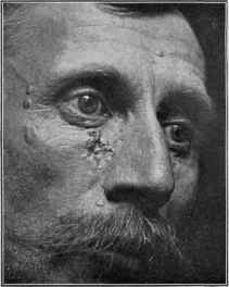
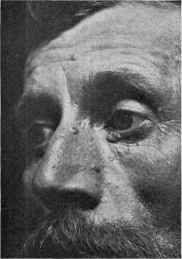
Fig. 154.—Multiple benign cystic epithelioma; the coalescent group showing degen
erative changes.
pinkish, pearly, or pale yellowish, occasionally it is that of the normal
skin, and in some instances there is a bluish tinge. They are firmly
imbedded. Their growth is slow, and, after reaching the size of a small
or large pea, remain stationary. It has been commonly thought that
no degenerative or ulcerative changes ever take place, and this absence
of malignancy is the rule, but in White’s case, in one observed by Jarisch,1
and also in mine, the large or bunched lesions exhibited surface degenera
tion with ulceration, and approached closely to the rolled, pearly-bordered,
superficial epitheliomata. The growths show no tendency to involution2
While sometimes presenting a pseudovesicular appearance, they are
usually firm and apparently solid in character, and if pricked, show, with
1 Jarisch, Hautkrankheiten, 1900, p. 788.
2 Lesions in Ormsby’s case underwent involution, and in Dyer’s case there was a,
tendency to “self-destruction and selfelimination.”
654
NEW GROWTHS
few reported exceptions (Dyer, Perry), no liquid contents, but simply
bleed slightly.
The face is the most common site, and here there usually is a pre
dilection shown for the region of the eyelids, forehead, cheeks, root of
the nose, chin, ears, and interpalpebral space. The interscapular
region, breast, and arms are also not uncommon sites, and the lesions
have, moreover, been found on other regions; Ormsby’s exceptional
case was more or less generalized. There are no subjective symptoms,
nor is there any disturbance of the general health, the cases coming chiefly
under observation owing to the disfigurement produced.
Etiology and Pathology.—The cause of the disease is un
known. Both sexes are liable, and almost any age, although it usu
ally has its beginning during adolescence, and is much more common in
females. Brooke’s 3 cases consisted of mother and two daughters, and
Fordyce’s, of a mother and daughter, with the history in the latter
instance of a similar condition in a preceding generation. Colcott
Fox1 also noted probable examples, clinically viewed, of the malady
in mother and daughter. Quinquaud’s patient stated that a sister
presented similar growths.
Crocker,2 in a recent valuable contribution, expresses the opinion
that the cases reported really constitute two distinct types or affec
tions, for which he suggests, for the sake of convenience, of holding
to the names given to the representative of the one class by Kaposi—
lymphangioma tuberosum multiplex—and, to the other class, that by
Brooke, acanthoma adenoides cysticum; including, in the former,
without necessarily implying that the condition is lymphangiomatous,
the cases of Kaposi, Jacquet and Darier, Török, Quinquaud, Lesser
and Beneke, and others,3 and, in the latter, those of Perry, Brooke,
Fordyce, White, and some others. It is true that a study of these
various cases suggests some clinical, although probably unimportant,
differences. Crocker gives the chief of these: in lymphangioma tubero-
sum—mainly on the trunk, discrete and not grouped, bilateral and not
symmetric, distinctly colored, in males and females alike, and not
hereditary; and for acanthoma adenoides cysticum—mainly on face,
discrete, but very closely grouped, closely symmetric, almost or quite
pearly white, or a faint bluish or yellowish tinge, most of them hereditary,
and all females. Jarisch’s case and my case, however, which come in
this latter group, were males. To these Crocker would add anatomic
dissimilarity—the former consisting of “cysts in the derma, with straight
processes of non-epidermic origin,” and the latter, “solid, coil-like masses
with small cysts scattered through them and of epidermic origin.” Hart-
1 Colcott Fox, Brit. Jour. Derm., 1897, p. 230 (case demonstration).
2 Crocker, “A Case of Lymphangioma Tuberosum Multiplex,” London Clin. Soc’y
Trans., 1899, vol. xxxii, p. 151 (with colored plate and bibliography); Sutton and
Dennie, “Possible Interrelationship of Acanthoma Adenoides Cysticum (Multiple
Benign Cystic Epithelioma) and Syringocystadenoma (Lymphangioma Tuberosum
Multiplex). Jour. Amer. Med. Assoc, Feb. 3, 1912, p. 333 (discuss the subject, and
record two cases, each representing distinct groups; review and references).
3 Literature references to the cases of Kaposi, Lesser, and Beneke, and also to Hog-
gan’s and Jarisch’s papers, which concern cases of benign cystic epithelioma, etc, are
given under lymphangioma.
MULTIPLE BENIGN CYSTIC EPITHELIOMA 655
zell and Heidingsfeld believe that these (or most of these) variously named
cases are simply varieties of the one and same affection, while C. J.
White contends that there are several distinct clinical and pathologic
groups.
Histologically (Darier, Brooke, Fordyce, Bowen, and others) the
lesion is shown to be an epithelial growth, being constituted of
irregularly rounded, oval, and elongated masses and tracts of epithe
lial cells corresponding to those in the lowermost layer of the
epidermis and external root-sheath of the hair-follicle; these masses
being distinct or composed of intercommunicating bands and tracts,
in some places resembling coil ducts; cell-nests are to be seen, as in
malignant epithelioma (Fordyce). Colloid degeneration is also noted.
Lying in the tracts, or more generally in the masses, were cysts of circular
or oval shape, sometimes elementary, others well formed, filled with either
purely colloid matter or partly with colloid and partly with concentric
layers of apparently horned epithelium (Brooke). It is generally believed
(Quinquaud, Jacquet, Darier, Philippson, Fordyce) that the growths
take their start from embryonic epithelial germs misplaced during fetal
life, and remaining in a latent condition until excited by some influence
into active proliferation (Fordyce), and this excitation is apparently
furnished most frequently at the period of puberty, doubtless by the
tissue changes and glandular activity at this time of life. In Hartzell’s
cases the growth had its origin in the epithelium of the hair-follicle, and
C. J. White’s investigation showed his to be a new growth and cystic
dilation of sweat ducts. Its relation to superficial epithelioma or rodent
ulcer is probably a close one, and although the lesions are thought benign
and to show no destructive changes, the exceptional cases of White,
Jarisch, and my own furnish, in my judgment, connecting examples.1
Philippson,2 in his report of a case, has endeavored to show that colloid
degeneration of the skin and benign cystic epithelioma are essentially
pathologically identical, a view, however, that has received no support.
Diagnosis.—The lesions bear some resemblance to molluscum
contagiosum, but are distinguished from the latter by the fact that
they are persistent, showing no tendency to disappear, and have, as a
rule, no central depression, and have no central aperture. Molluscum
1 Adamson, Lancet, Oct. 17, 1908, in an interesting analytical and critical paper,
discusses this question. He considers that the Jarisch, White, and Stelwagon (mine)
cases are closer to the rare examples of multiple rodent ulcer (2 cases cited and pictured)
than to the true (Brooke) type of multiple benign cystic epithelioma, the latter clin
ically differing in these particulars; (1) all have occurred in women, and the lesions
appeared in childhood; (2) generally in mother and daughter; (3) distribution of lesions
markedly symmetrical; (4) fairly uniform size of lesions; (5) no tendency of the lesions
to enlarge beyond the size of a split pea, nor to break down, i. e., to become locally
malignant. Although the writer would evidently like to hold to the distinct indi
viduality of the several groups, he recognizes, however, that White’s case might be
looked upon as a connecting link; and that, histologically, the lesions of rodent ulcer
have many features in common with those of multiple benign cystic epithelioma; and
that, following the cases in series, the difference does not seem so great. The paper
leads to the final query that has not yet been answered—“as to what is the essential
difference between a benign and a malignant epitheliomatous growth.”
2 Philippson, “Die Beziehungen des Kolloid-milium (Wagner) der kolloiden De
generation der Cutis (Besnier) und des Hydradenom (Darier-Jacquet) zu Einander,”
Monatshefte, 1890, vol. xi, p. 1; and also in Brit. Jour. Derm., 1891, p. 35.
656
NEW GROWTHS
contagiosum is, moreover, more commonly a malady of childhood:
benign cystic epithelioma rarely presents before puberty. There is
also a resemblance to hydrocystoma, but in the latter the growths
have fluid contents and are usually fluctuating as to their existence,
disappearing and reappearing. Between colloid degeneration of the
skin and this disease there is also some clinical similarity. The colloid
lesion begins as yellow, translucent, gelatinous-looking nodule: cys
tic epithelioma as a small, skin-colored papule or black dot, and
gradually progresses; the former may undergo involution and disap
pear without trace; the latter is persistent. Histologically in the for
mer the colloid material is infiltrated in the fibrillæ of the connective
tissue, enveloping the connective tissue bundles and following their
directions, and there are no epithelial tracts or cords, and no cysts—
findings different from those of cystic epithelioma (Brooke).
Prognosis and Treatment.—There is no tendency to sponta
neous disappearance, and though the malady is usually to be considered
benign, development of a more active epithelial proliferation and ulcera-
tive degeneration is a possibility. In view of the epitheliomatous
development in his case White justly says, I believe, that the correctness
of the appellation benign must be regarded as problematic. Treatment
is surgical. Fordyce has found that simple incision in the smaller
lesions and squeezing out the growth will sometimes be successful,
although stating that curetting constitutes the best plan. In a case un
der the care of Dr. C. N. Davis, of Philadelphia, that I had an opportu
nity of seeing, in spite of several thorough curettings there was per
sistent recurring tendency. Electrolysis and cauterization can also be
resorted to. In Ormsby’s case x-ray treatment and carbon-dioxid snow
proved of value. The method by curetting and supplementary cauter
ization seems to me the best.
But first, if you want to come back to this web site again, just add it to your bookmarks or favorites now! Then you'll find it easy!
Also, please consider sharing our helpful website with your online friends.
BELOW ARE OUR OTHER HEALTH WEB SITES: |
Copyright © 2000-present Donald Urquhart. All Rights Reserved. All universal rights reserved. Designated trademarks and brands are the property of their respective owners. Use of this Web site constitutes acceptance of our legal disclaimer. | Contact Us | Privacy Policy | About Us |