| MEDICAL INTRO |
| BOOKS ON OLD MEDICAL TREATMENTS AND REMEDIES |
THE PRACTICAL |
ALCOHOL AND THE HUMAN BODY In fact alcohol was known to be a poison, and considered quite dangerous. Something modern medicine now agrees with. This was known circa 1907. A very impressive scientific book on the subject. |
DISEASES OF THE SKIN is a massive book on skin diseases from 1914. Don't be feint hearted though, it's loaded with photos that I found disturbing. |
PAGET’S DISEASE
Synonyms.—Paget’s disease of the nipple; Malignant papillary dermatitis (Thin);
Eczema epitheliomatosa; Eczematoid epitheliomatosis of the nipple; Cutaneous psoro-
spermosis; Psorospermosis cutis; Mammillaris maligna; Fr., Maladie de Paget; Epithé-
liome de Paget; Ger., Paget’s Krankheit.
Definition.—Paget’s disease is a rare malignant disease, usually
of the nipple and areola in women, beginning as an inflammatory-looking,
eczematoid affection, and eventually terminating in cancerous involve
ment of the whole gland.
Attention was first called to this malady by Paget1 in 1874, whose
description was based upon an observation of 15 cases, in all of which
—women between the ages of forty and sixty—cancerous involvement
of the gland followed within one or two years after the appearance of
the cutaneous symptoms. Since then many additional cases have been
reported, and the malady has received considerable attention, both in
its clinical and histopathologic aspects, by various observers,2 among
whom Butlin, Thin, Duhring, Wickham, Bowlby, Hutchinson, Jr.,
Jackson, Wiggin and Fordyce, Hartzell, Simpson, and others.
Symptoms.—The disease is exceedingly insidious in its appear
ance, and scarcely comes under notice until a distinctly eczematoid
aspect is presented. In its very earliest stage it consists of slight, scaly,
somewhat hardened, thin, epidermic collections or scurnness of the
nipple and the immediately contiguous portion of the areola, with,
1 Paget, St. Bartholomew’s Hospital Reps., 1874, vol. v, p. 87.
2 Butlin, London Med.-Chirurg. Soc’y Trans., 1876, vol. lix, p. 107, and 1877, vol.
lx, P. 153 (with histologic illustrations); Thin, London Patholog. Soc’y Trans., 1881,
vol. xxxii, p. 218 (with histologic cuts), and Brit. Med. Jour., 1881, vol. i, pp. 760, 798
(with histologic cuts), “On Cancerous Affection of the Skin,” London, 1886 (with review
of the subject); Dunring and Wile, Amer. Jour. Med. Sci., 1884, vol. lxxxviii, p. 141
(pathology with references); Wickham, “Maladie de la peau dite maladie de Paget,”
Thèse de Paris, 1890 (with colored plates, review, and bibliography); and Annales,
1890, pp. 45 and 139 (with bibliography); Bowlby, London Med.-Chirurg. Soc’y
Trans., 1891, p. 341 (notes of 13 cases); Hutchinson, Jr., London Patholog. Soc’y Trans.,
1890, p. 214 (with histologic cuts), Brit. Jour. Derm., 1891, p. 278; G. T. Jackson,
Jour. Cutan. Dis., 1896, p. 428 (with review and important references); Wiggin and
Fordyce, New York Med. Jour., 1897, vol. 1xvi, p. 445 (with colored case illustration
and histologic cuts); Hartzell, Jour. Cutan. Dis., 1906, p. 289 (2 cases, x-ray treatment,
with report of microscopic findings in one of them after prolonged treatment); Simpson,
Quar. Bull. Northwest Univ. Med. School, June, 1909 (case, histology, review, and ref
erences; decidedly benefited by x-rays).
CARCINOMA CUTIS
867
later, slight redness and often more or less itching. It may remain
limited to this small circumscribed area for months or longer, during
which time slight or moderate erosion of the nipple may present and
crusting ensue. After a variable time the condition spreads out and
soon involves the whole area of the areola, and often extends beyond.
When at all developed, the diseased area, which is usually sharply mar-
ginate, exhibits a florid, intensely red, very finely granular, raw surface,
attended with a more or less viscid exudation. There is moderate in
filtration, which is well defined below, feeling, in fact, like a thin layer of
indurated tissue implanted in the skin.
The malady slowly progresses, fissuring, erosion, and retraction of
the nipple gradually ensuing, which sooner or later has entirely disap
peared. After some months or several years the process becomes more
intense, greater thickening is noted, the nipple and contiguous part of
the areola are ulcerated or have “melted away,'’ and some nodular
hardening usually develops in the gland structure—in short, gradual
scirrhous involvement of the whole breast finally occurs. As a rule, the
superficial or eczematoid area does not extend more than several inches
beyond the areola, but in some instances, as notably in those reported
by Jamieson1 and Elliot,2 it is much more extensive; in these 2 cases
the entire surface of the breast was involved and the axillary region partly
invaded. In a few instances, too, the malady has affected both breasts.
The course of the malady is, moreover, extremely variable. In some cases,
as in those reported by Paget, but one or two years elapsed before cár-
cinomatous development in the gland was noted; in others the disease
remains for a long time confined to the surface as an eczematoid eruption
—in Morris’s3 case six years, in Duhring’s case ten years, and in Jamie-
son’s twenty years. As a rule, however, in two or three years malignant
involvement of the breast has ensued.
According to the observations of recent years, it would seem that
the disease is not necessarily one limited to the breast. Crocker4 has
observed an instance of its occurrence on the scrotum, Tommasoli5
on penis, Pick6 on the glans penis, Sheild7 on pubic region, extending
on to penis and scrotum, Dubreuilh8 on the vulva, Darier and Couil-
laud9 on the scrotum and perineal region, Winfield10 on the lip, and
Ravogli11 on the nose; Jungmann and Pollitzer12 in the axilla, Colcott
1 Jamieson, Diseases of the Skin, p. 482 (woman aged seventy two).
2 Elliot, Jour. Cutan. Dis., 1892, p. 272.
3 Henry Morris, London Med.-Chirurg. Soc’y Trans., 1880, vol. lxiii, p. 37 (colored
plate case illustration and histologic cuts).
4 Crocker, London Patholog. Soc’y Trans., 1889, vol. xl, p. 187 (with colored plate
case illustration and histologic cuts).
5 Tommasoli, Giorn. ital., 1893, vol. xxviii, Fasc iv.
6 Pick, Prager. med. Wochenschr., 1891, p. 282.
7 Sheild, Brit. Jour. Derm., 1897, p. 35 (man aged sixty).
8 Dubreuilh, ibid., 1901, p. 407.
9 Darier and Couillaud, Annales, 1893, p. 33 (man aged seventy-two, fifteen years’
duration).
10 Winfield, Brooklyn Med. Jour., March, 1896 (Soc’y proceedings).
11 Ravogli, Trans. Internat. Med. Cong., Rome, 1894; abs. in Jour. Cutan. Dis., 1894,
p. 222 (patient an old lady).
12 Jungmann and Pollitzer, Dermatolog. Zeitschr., June, 1904.
868
NEW GROWTHS
Fox and Macleod1 in the umbilical region, Fordyce,2 probable case
on the buttocks, Davis3 on the penis, and Hartzell4 on the forearm.
About 18 extramammary cases are a matter of record, and of those
9 occurred on the external genitalia (Hartzell). I have met with a
case somewhat similar to Ravogli’s case, in a woman aged sixty, the
whole nose being superficially involved and eroded and clinically sug
gestive of this malady. An instance of its occurrence on the scrotum
has also come under my notice in an old man (Dr. C. N. Davis’ patient,
not elsewhere recorded).
Etiology.—The disease is one of advancing years, occurring
most frequently between fifty and sixty. It is practically limited to
the female sex and to the nipple region, the cases occurring on other
parts in men still being viewed with some suspicion. In one instance,
observed by Forrest,5 however, of apparently eczematous disease of the
nipple in a male aged seventy-two, carcinoma developed. There is a
somewhat remarkable disproportion in its occurrence on the right side;
in not more than 25 per cent, was the left breast the seat of the disease.
Various causes have been considered as etiologic. The malady was
formerly thought to be a carcinoma developing upon a long-continued
eczema, but it is now generally believed that the process is malignant
from the start. Doubtless fissures and persistent irritation of the nipple
are favoring factors. Darier and Wickham advanced the opinion that
psorosperms are the exciting agents; psorosperm-like bodies have also
been found by Bowlby, Macallum,6 Hutchinson, Jr., and others. This
view is, however, no longer maintained; that originally held by Thin,
and later by Unna, Fordyce, and others, that these bodies merely repre
sent cell changes, is now generally accepted.7
Pathology.—At the present time there is but little doubt as to
the malignant nature of even the earliest phases of the malady.
The pathologic anatomy has been studied by various observers
(Butlin, Thin, Duhring, Darier, Wickham, Fordyce, Unna, Hartzell,
and others). There is practically more or less unanimity in the findings.
“The morbid changes (quoting Fordyce) may be briefly stated as in
flammation of the papillary region of the derma, leading to an edema and
vacuolation of the constituent cells of the epidermis, followed by their
complete destruction in places and their abnormal proliferation in others.
The change in the epithelium of the lactiferous canals and glandular
1 Colcott Fox and MacLeod, Brit. Jour. Derm., 1904, p. 43 (with case illustration,
histologic cuts, review of these special cases and a general review of the disease, and
references; man aged sixty-five, of eleven years’ duration).
2 Fordyce, Jour. Cutan. Dis., 1905, p. 193 (with histologic cuts), a probable case of
the gluteal region (woman, aged sixty, of six years’ duration).
3 C. N. Davis, Jour. Cutan. Dis., 1910, p. 412 (case demonstration).
4 Hartzell, “Extramammary Paget’s Disease,” Jour. Cutan. Dis., 1910, p. 379
(report of case on forearm, refers to 4 unpublished cases; review, and bibliography;
case and histologic illustrations).
5 Forrest, Glasgow Med. Jour., vol. xvi, p, 459 (patient aged seventy-two).
6Macallum, Canadian Med. Practitioner, 1890, p. 473.
7 Fabry and Trautmann, Archiv, 1904, vol. lxix, p. 37, found an yeast fungus, and
suggest a possible relationship between Paget’s disease and blastomycetic dermatitis.
Inasmuch as this has not been observed by other careful investigators, it is probable
that in this instance its presence was secondary or accidental.
CARCINOMA CUTIS 869
epithelium, which is also of a proliferative and degenerative nature, is
secondary to the changes in the surface epithelium, and may be regarded
as of the same nature, and probably produced by the action of the same
irritant. The over-distention of the lactiferous canals by the proliferat
ing epithelium, resulting in a malignant infection of the surrounding
connective tissue, is the usual termination of the affection.’’ As all
observers have found, as Fordyce further states, “the earliest and most
carefully studied changes in Paget’s disease are those met with in the
surface epithelium. It is here that the cell changes and inclusions are
met with which were first described by Darier, and afterward by Wick-
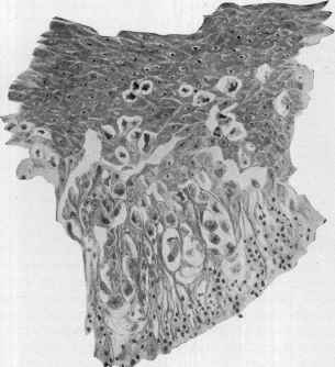
ham and others, as coccidia. ... A more careful study of these

Fig. 211.—Paget’s disease, in middle stage, showing the peculiar epithelial cell degener
ation and the psorosperm-like bodies (courtesy of Dr. A. R. Robinson).
cell degenerations has pretty conclusively demonstrated the non-parasitic
character of many of them. The infectious nature of Paget’s disease
has, however, by no means been absolutely disproved, and an element
of doubt yet remains as to the character of certain of the cell changes
which are found in the affection.” The rôle which Darier, Wickham,
and others gave these peculiar cell-degenerations, under the erroneous
impression that they were coccidia or psorosperms, has, as already stated,
been practically abandoned.
Diagnosis.—The disease, in its earliest stage, is to be distin
guished from eczema, a matter in some instances of some difficulty,
until the case has been under observation for a short period. In its
870
NEW GROWTHS
later stages, and especially when the gland involvement is already evi
dent, a mistake could occur only as a result of a hasty and careless exami
nation. The diagnostic features are: The age of the patient; the sharp
limitation; the well-defined, indurated film of infiltration; the peculiar,
red, raw, granulating appearance; and, later, the retraction of the nipple;
and, finally, the involvement of the deeper parts. A persistent circum
scribed eczematous-looking eruption of the nipple and areola should
always be viewed with suspicion in those advancing in years, which be
comes almost a certainty if it is rebellious to the usual treatment of
eczema. In the earlier stage in doubtful cases examination may be
made for the psorosperm-like bodies, which are characteristic, and not
to be found in eczema.
Prognosis and Treatment—If the disease is recognized early
and properly treated a cure may be often anticipated; but later the
prognosis is essentially the same as that of scirrhus of the breast, and
depending upon the progress of the disease and the amount of breast
involvement.
Treatment, when the diagnosis is clearly established, should be
radical, consisting of the plans mentioned for epithelioma, radical opera
tion being the first choice. While mild and palliative applications can
do no direct harm, half-way measures are not permissible, as the latter
simply serve to spur the disease to more rapid advancement. In doubt
ful cases the various plans of treating eczema are at first to be employed,
and if this disease, the condition will usually readily yield. In clear
cases of the disease, in which radical measures are refused, palliative and
soothing applications are to be made. In Elliot’s patient the use of an
ointment of fuchsin, 2 to 5 grains (o. 135-0.35) to the ounce (32.) of
lanolin and rose-water, of a strength just short of producing irritation,
acted satisfactorily, giving considerable relief and promoting cicatrization.
X-ray treatment sometimes benefits, and in the very earliest stage
of the disease, before the ducts and glands are involved, might prove
curative; 1 of Hartzell’s cases, 1 of my cases, and Simpson’s case improved
under this; Jungmann and Pollitzer report a cure of their axilla case and
Fordyce in his gluteal case; and Milligan a cure of umbilicus case with
radium.1
But first, if you want to come back to this web site again, just add it to your bookmarks or favorites now! Then you'll find it easy!
Also, please consider sharing our helpful website with your online friends.
BELOW ARE OUR OTHER HEALTH WEB SITES: |
Copyright © 2000-present Donald Urquhart. All Rights Reserved. All universal rights reserved. Designated trademarks and brands are the property of their respective owners. Use of this Web site constitutes acceptance of our legal disclaimer. | Contact Us | Privacy Policy | About Us |